
Category: Dermatology

Dermatology issues can affect comfort, sleep, and social confidence daily. This category helps patients and caregivers review common skin, hair, and nail concerns. It also supports US delivery from Canada for eligible prescription refills. Use the links to learn terms, track symptoms, and plan next steps.

Topics range from acne treatment and eczema care to psoriasis management. Some posts cover screening basics, like mole checks and melanoma awareness. Others explain tests such as patch testing (allergy checks for contact triggers). Dispensing is handled by licensed Canadian partner pharmacies, when medication is supplied.

Dermatology Resources and Navigation

Start with the educational posts if a diagnosis feels unclear. Many guides explain how clinicians describe rashes, plaques, and flares. For autoimmune patterns, see the Everything To Know Autoimmune Diseases overview.

Use the product listings when a prescription name is already known. Each page summarizes form, label directions, and typical documentation needs. Condition campaigns can also help families talk about long-term skin care. Browse World Psoriasis Day 2025 for community context and support planning.

What You’ll Find in This Category

This hub gathers practical guides on common skin conditions and evaluations. Readers will see plain-language explanations alongside clinical terms used in clinics. Common topics include rosacea, dermatitis evaluation, and hives and urticaria (itchy welts).

Dermatology content here also connects to related prescription options below. Browse the Skin Health Product Category to compare available listings. Some entries discuss therapies often used for psoriasis, including Otezla and Apremilast. Related reading includes Otezla Uses and Apremilast Mechanism Of Action. For biologic options, see Cosentyx and the Exploring Cosentyx Uses guide.

  • Acne, blackheads, and scarring concerns
  • Eczema and atopic dermatitis flare patterns
  • Psoriasis types, triggers, and long-term planning
  • Rosacea redness, bumps, and eye irritation questions
  • Rash diagnosis basics, including contact triggers and exposures
  • Warts, including common options for wart removal
  • Hair and nail disorders, including alopecia (hair loss) topics
  • Nail fungus treatment basics and fungal skin infections
  • Phototherapy overviews and what to ask a clinic
  • Skin cancer screening terms, including melanoma screening
  • Mole check guidance and what changes clinicians track
  • Actinic keratosis (rough, sun-damaged spot) explanations

How to Choose

Choosing a topic path can feel overwhelming, especially during flares. Dermatology care can look different for children, adults, and older relatives. Start by separating symptom questions from treatment or access questions. Awareness posts, like Psoriasis Awareness Month, can support shared language.

Quick tip: Keep a dated photo log for rashes, nails, or scalp changes.

Use a simple comparison checklist

Use this checklist to narrow what to read next. It also helps caregivers share clearer details with clinicians.

  • Where symptoms appear and whether they move or spread
  • How long symptoms last and how often they return
  • Main sensation, like itch, pain, burning, or tightness
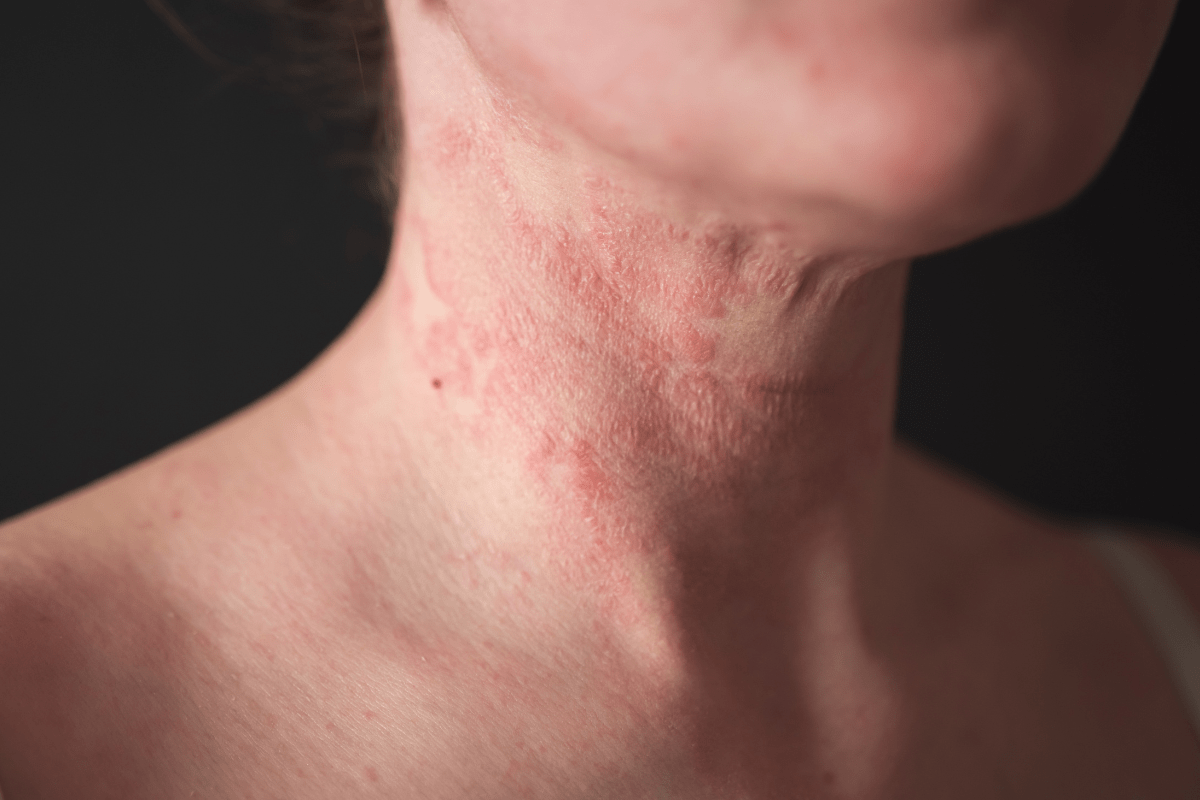
  • Visible pattern, like ring-shaped, scaly, or blistering spots
  • Recent exposures, including new soaps, metals, plants, or pets
  • New medicines or supplements started before symptoms appeared
  • Age group, including infant, child, teen, or older adult needs
  • Other health issues, especially immune conditions and diabetes
  • Prior treatments tried and what happened afterward
  • Preference limits, like scent-free products or injection concerns
  • Testing needs, such as patch testing or lab monitoring
  • Visit format, including in-person exams or teledermatology follow-ups

Know when a specialist evaluation adds value

A primary care visit can address many common skin problems. A skin specialist can help when the diagnosis stays uncertain. They may also help with persistent scalp, nail, or genital involvement. Families often benefit from written plans that reduce confusion at home.

Safety and Use Notes

Safety matters because skin findings sometimes signal broader health issues. Dermatology information can help frame questions for a clinician visit. Some symptoms need prompt evaluation, such as fast-spreading rashes or fever. For skin self-check basics, review the AAD Skin Self-Check Guide.

Many skin medicines can irritate, thin, or lighten skin with misuse. Topical steroids, retinoids, and antifungals all carry label precautions. Phototherapy can cause burns without careful dosing and follow-up. When required, prescriptions are verified directly with the prescriber. For a plain-language psoriasis overview, see the MedlinePlus Psoriasis Overview.

  • Share allergy history, including reactions to adhesives or topical antibiotics
  • Avoid sharing prescriptions, even with similar-looking rashes
  • Ask about interactions if using multiple creams on the same area
  • Note pregnancy, breastfeeding, or fertility plans before starting new medicines
  • Report new infection signs when using immune-modifying treatments
  • Track lesion changes, including size, color, bleeding, or crusting
  • Use sun protection discussions to support long-term skin cancer prevention
  • Keep label instructions handy to reduce missed or doubled doses

Access and Prescription Requirements

Some pages in this hub link to prescription product details and related education. Many medications require a valid prescription from a licensed prescriber. The process may involve confirming prescriber contact information and current directions. This helps reduce avoidable delays and incorrect dispensing.

Cash-pay options can help, including for people without insurance. For some items, refills depend on prescriber authorization and local rules. Keep names, birthdates, and addresses consistent across documents. Use your account tools to store needed details for future requests.

Why it matters: Clear paperwork can reduce back-and-forth when prescriptions must be confirmed.

  • Have prescriber details available for verification when an Rx is required
  • Check that directions and quantity match what the prescriber intended
  • Plan ahead for items that need refrigeration or special handling
  • Review return limits for prescriptions, since many are not returnable
  • Keep a current medication list to support safe pharmacy review
  • Ask the prescriber about monitoring needs tied to the prescribed therapy

This content is for informational purposes only and is not a substitute for professional medical advice.

  • Otezla Uses in Psoriasis Care: Safety and Key Questions

    Otezla Uses in Psoriasis Care: Safety and Key Questions

    Otezla uses include treating certain inflammatory conditions, mainly plaque psoriasis, psoriatic arthritis, and oral ulcers linked to Behçet’s disease. It is an oral prescription medicine, so people often consider it when they want a non-injectable systemic option. Why this matters: knowing what it treats, how it differs from biologics, and which side effects need attention can make treatment discussions more focused.

    This article explains Otezla (apremilast) in plain language. It covers approved uses, how the drug works, common side effects, weight and hair questions, alcohol concerns, and when to recheck with your clinician.

    Key Takeaways

    • Approved uses: Plaque psoriasis, psoriatic arthritis, and Behçet’s-related oral ulcers.
    • Oral treatment: It is a tablet, not an injection or infusion.
    • Not a biologic: It is a targeted small-molecule medicine.
    • Common effects: Diarrhea, nausea, headache, and appetite changes may occur.
    • Safety focus: Mood changes and unintended weight loss deserve prompt attention.

    Where Otezla Fits in Plaque Psoriasis and Joint Care

    Otezla uses are tied to inflammation in the skin, joints, and mouth. In plaque psoriasis, clinicians may consider it when topical treatments are not enough, or when a systemic treatment is appropriate. In psoriatic arthritis, the goal is often to reduce joint pain, stiffness, swelling, and related limitations. For Behçet’s disease, it is used for painful oral ulcers in adults.

    Psoriasis is more than a skin surface problem. It involves immune signaling that can affect the skin, nails, joints, and overall quality of life. Some people mainly want less itch or scaling. Others care most about hand function, morning stiffness, sleep, or avoiding injections. Your main treatment goal matters because psoriasis medicines differ in route, monitoring, safety profile, and expected role in care.

    If you are reviewing medication names, the product summary for Otezla can help you recognize the brand while you discuss options with your prescriber. For broader category browsing, the Dermatology Products collection can help you compare general treatment routes without replacing clinical advice.

    Why it matters: A clear treatment goal helps your clinician compare realistic options.

    How Apremilast Works, and What It Is Not

    Apremilast is the generic name for Otezla. It works by inhibiting phosphodiesterase 4, often shortened to PDE4. PDE4 is an enzyme involved in inflammatory signaling inside immune cells. By affecting that pathway, apremilast may help reduce some inflammatory signals linked to psoriasis and psoriatic arthritis.

    Many people ask, is Otezla a biologic. It is not. Biologics are large protein-based medicines, usually given by injection or infusion. Apremilast is a small-molecule tablet. That difference can affect storage, route, and monitoring conversations, though it does not automatically make one option better for every person.

    Another common question is whether it is a steroid. It is not a corticosteroid. It also is not the same as methotrexate, even though both may be discussed as systemic options. Some clinicians may refer to apremilast as a targeted synthetic drug because it is an oral, non-biologic therapy that targets a specific intracellular pathway.

    For a deeper mechanism overview, read Apremilast Mechanism of Action. If you are comparing autoimmune conditions more broadly, Autoimmune Diseases can help explain why immune-directed treatments differ across diagnoses.

    Starting Treatment: What to Expect Early On

    Otezla is taken by mouth, usually as a tablet. Many people start with a short titration schedule, meaning the dose is increased gradually. This step-up approach is designed to help with early tolerability, especially stomach-related symptoms. Your prescriber decides the schedule and whether any adjustment is needed for your situation.

    Symptom improvement is not always immediate. Skin plaques, itch, nail changes, and joint symptoms may shift at different speeds. A weekly symptom note can help you see patterns instead of reacting to one difficult day. Track the issues that actually affect your life, such as itch, pain, sleep, hand function, or missed activities.

    Kidney function can matter for dosing in severe impairment. Tell your clinician if you have kidney disease, dialysis, or recent major lab changes. Also share your full medication list, including supplements. Certain medicines can affect how apremilast is handled in the body.

    For people comparing generic and brand naming, the Apremilast listing can support medication-name discussions with a pharmacist or prescriber. BorderFreeHealth connects U.S. patients with licensed Canadian partner pharmacies, and prescription details are verified when required before pharmacy dispensing.

    Otezla Side Effects: Common, Serious, and Worth Tracking

    Otezla side effects most often involve the stomach or nervous system early in treatment. Diarrhea, nausea, abdominal discomfort, headache, and reduced appetite are commonly reported. Some people also notice upper respiratory symptoms, though those can be hard to separate from routine viral illnesses.

    Some early side effects may ease as the body adjusts, but that is not guaranteed. Hydration, taking the medicine consistently as prescribed, and following the titration plan may help some people tolerate treatment. If symptoms interfere with eating, drinking, work, or sleep, contact your prescriber rather than forcing yourself through it.

    Serious or persistent symptoms deserve faster attention. These include severe diarrhea, ongoing vomiting, signs of dehydration, faintness, or symptoms that stop you from keeping fluids down. New or worsening depression, unusual mood changes, or thoughts of self-harm need urgent support.

    Otezla uses and side effects should be discussed together, not separately. A medicine may fit well for one person and poorly for another based on mental health history, weight trends, kidney function, other medications, and tolerance for stomach symptoms. People who read online reviews should remember that forums often overrepresent very strong experiences.

    Weight changes

    Otezla weight loss is specifically noted in prescribing information. It does not happen to everyone, but unintended weight loss should be taken seriously. Reduced appetite, nausea, diarrhea, or changes in eating patterns may contribute for some people. Others may notice weight changes without an obvious stomach trigger.

    A simple weight log can help you and your clinician judge whether a change is meaningful. If you are already underweight, have an eating disorder history, have a serious chronic illness, or are losing weight without trying, raise that early.

    This calculator can help track body-weight change as a general number, but it cannot judge whether a medication is safe for you.

    [weight-loss-progress-calculator]

    Hair and body changes

    People also ask, does Otezla cause hair loss. Hair loss is not usually highlighted as one of the most common labeled side effects, but individual experiences vary. Psoriasis flares, scalp inflammation, stress, thyroid disease, iron deficiency, and other medicines can also affect shedding. A clinician can help check common causes before assuming one drug is responsible.

    Alcohol, Vaccines, Infections, and Daily-Life Cautions

    Alcohol is not known as a major direct interaction for most people taking apremilast, but it can worsen dehydration and stomach irritation. That overlap matters if diarrhea, nausea, or poor appetite are already present. It can also affect mood and sleep, which may complicate monitoring.

    Otezla is not a classic broad immunosuppressant in the same way as some older systemic medicines, and it is not a steroid. Still, it influences immune signaling. Tell your clinician about frequent infections, serious recent illness, or other immune-directed medicines. Vaccine timing depends on your overall health and the specific vaccine.

    Some readers ask whether Otezla is a high-risk medication. Risk depends on context. For many people, clinicians focus on tolerability, mood, weight, medication interactions, and kidney function. It may not require the same routine lab monitoring as some other systemic therapies, but that does not mean follow-up can be skipped.

    Quick tip: Bring a current medication list to every dermatology or rheumatology visit.

    People without insurance sometimes compare cash-pay prescription options across countries. BorderFreeHealth supports access to cross-border prescription options for eligible patients, subject to jurisdiction and prescriber requirements, but clinical suitability still belongs in a clinician-led discussion.

    How It Compares With Other Psoriasis Treatment Paths

    Apremilast sits between several common psoriasis care paths. Topical medicines act mainly on the skin surface. Phototherapy uses controlled light exposure. Conventional systemic medicines, such as methotrexate, affect broader immune pathways and often require lab monitoring. Biologics target specific immune proteins and are usually injected or infused.

    The right comparison depends on your main problem. A person with mostly skin plaques may ask different questions than someone with swollen fingers, heel pain, or morning stiffness. Nail disease, scalp involvement, pregnancy plans, infection history, mood history, and kidney function can also shape the discussion.

    Otezla cost questions are common, including Medicare or monthly-cost concerns. Exact costs vary by location, coverage, pharmacy, assistance programs, and product version. Because prices can change, it is safer to confirm directly with the pharmacy, insurer, or care team rather than relying on old online figures. If you are comparing newer oral dermatology options, the Sotyktu product page can help you identify another medication name to discuss, without implying it is interchangeable.

    A practical appointment question is: “What are we trying to improve first?” The answer may be clearer skin, less itch, better joint function, fewer flares, or lower treatment burden. Once that goal is named, side effects and monitoring become easier to weigh.

    When to Recheck With Your Clinician

    Recheck sooner if side effects feel severe, unusual, or unsafe. Do not wait for a routine appointment if you have persistent vomiting or diarrhea, symptoms of dehydration, major unintended weight loss, or new mood symptoms. Urgent mental health symptoms need urgent help.

    Schedule a planned follow-up if benefits are unclear after the trial period your prescriber recommended. Bring your symptom notes, weight trends, and any missed doses. If you started or stopped another medicine, developed a major illness, or are planning surgery, mention that too.

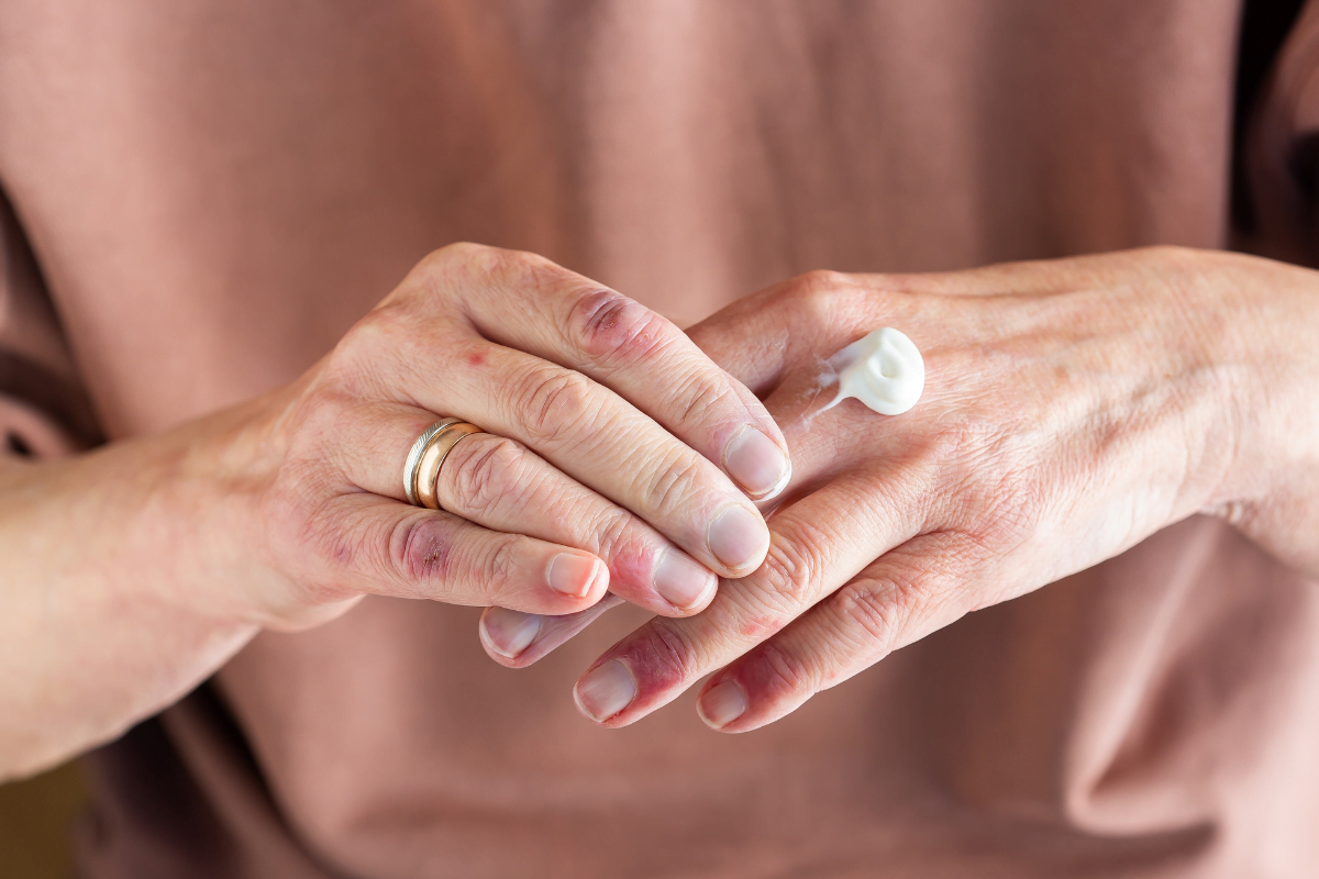
    Some people also ask whether petroleum jelly helps psoriasis. Plain petroleum jelly does not treat the immune cause of psoriasis, but it may help seal in moisture and reduce dryness or cracking for some plaques. Avoid applying it to infected, oozing, or uncertain rashes without medical guidance.

    For ongoing education, the Dermatology Articles collection can help you build a question list before appointments. Keep your list short and specific so the visit stays focused.

    Authoritative Sources

    For official indication, warning, and adverse-reaction details, review the manufacturer prescribing information for Otezla.

    For psoriasis background and treatment categories, the American Academy of Dermatology psoriasis overview offers patient-focused education.

    For vaccine timing and general immunization guidance, see the CDC vaccine information for patients.

    Recap

    Otezla uses include plaque psoriasis, psoriatic arthritis, and oral ulcers linked to Behçet’s disease. It is an oral targeted medicine, not a biologic and not a steroid. Common side effects include stomach symptoms and headache, while mood changes and unintended weight loss require careful attention.

    The best next step is a clear, practical conversation with your clinician. Ask what symptom you are treating first, what side effects should trigger a call, and how you will judge whether the medicine is helping.

    This content is for informational purposes only and is not a substitute for professional medical advice.

  • Apremilast Mechanism of Action: Uses, Safety, and Fit

    Apremilast Mechanism of Action: Uses, Safety, and Fit

    Apremilast mechanism of action centers on PDE4 inhibition, which changes inflammatory signaling inside immune cells. In plain terms, it helps turn down some immune messages linked with psoriasis, psoriatic arthritis, and Behçet’s-related mouth ulcers.

    That answer matters because apremilast is not a biologic injection, and it is not a steroid. It is an oral small-molecule medicine with a different monitoring and side-effect profile. Understanding that difference can help you ask clearer questions before starting or continuing treatment.

    Key Takeaways

    • Drug class: oral phosphodiesterase 4 inhibitor.
    • Main action: raises cAMP inside immune cells.
    • Common uses: plaque psoriasis, psoriatic arthritis, and Behçet’s oral ulcers.
    • Common effects: nausea, diarrhea, headache, and appetite changes.
    • Safety focus: mood changes, weight loss, kidney function, and interactions.

    How Apremilast Works Inside Immune Cells

    Apremilast works by inhibiting phosphodiesterase 4, usually shortened to PDE4. PDE4 is an enzyme found in several immune cells. Its job includes breaking down cyclic adenosine monophosphate, or cAMP, a messenger that helps regulate inflammatory activity inside cells.

    When PDE4 is blocked, cAMP levels rise. That shift can reduce the release of some pro-inflammatory cytokines (immune messenger proteins) and may increase some anti-inflammatory signals. The result is not a total immune shutdown. It is better described as immune modulation, meaning it adjusts inflammatory signaling rather than broadly suppressing every immune response.

    This is why the Apremilast mechanism of action is often described as intracellular. It works inside immune cells, not by binding one cytokine outside the cell. Biologic medicines often target specific immune proteins such as TNF, IL-17, or IL-23. Apremilast acts earlier in the signaling chain by changing the messenger balance that influences several inflammatory pathways.

    Why it matters: A medicine’s target can shape dosing, monitoring, side effects, and expectations.

    What class of drug is apremilast?

    Apremilast is an oral PDE4 inhibitor. It is also considered a targeted synthetic disease-modifying medicine in some rheumatology contexts. It is not a biologic, because biologics are large protein-based medicines made in living systems. It is not a steroid, because it does not act like cortisol or broadly mimic steroid hormones.

    That distinction helps when people compare treatment options. A pill can feel less intimidating than an injection, but the best fit still depends on the condition, symptom pattern, other medicines, and personal safety factors. If you want a broader immune-system primer, Autoimmune Diseases explains how immune misfires can affect different tissues.

    How This Mechanism Relates to Psoriasis and Joints

    Apremilast may help psoriasis and psoriatic arthritis because both involve immune-driven inflammation. Psoriasis is often most visible on the skin, but it is not only a skin-surface problem. Immune cells and cytokines contribute to plaques, scaling, redness, and itch. In psoriatic arthritis, inflammatory signaling can also affect joints, tendons, nails, and surrounding tissues.

    People sometimes ask which organ is linked to psoriasis. The skin is the most obvious organ involved, but psoriasis is a systemic inflammatory condition. That means inflammation may involve more than the visible plaques. Some people also develop joint symptoms, nail changes, or other health associations that need a broader care plan.

    In plaque psoriasis, treatment goals often include fewer plaques, less scaling, and reduced discomfort. In psoriatic arthritis, goals may include less joint pain, less morning stiffness, and better daily function. Apremilast does not work instantly. Symptom changes often build gradually, and your clinician may ask how skin, joints, weight, mood, and stomach tolerance are changing over time.

    For a condition-focused look at when an oral psoriasis option may be considered, see Otezla Uses. For skin-care awareness and support context, Psoriasis Action Month covers practical ways people engage with long-term skin health.

    Why response can differ between people

    The same diagnosis can behave differently from person to person. One person may have mostly skin plaques. Another may have mild skin symptoms but more joint stiffness. A third may have nail changes, fatigue, or symptoms that flare with stress, infection, or medication changes.

    Because the Apremilast mechanism of action affects several inflammatory mediators indirectly, response can vary. Some people may find the oral format and monitoring style fit well. Others may need a different pathway, especially when symptoms are severe, rapidly progressing, or not controlled enough. Those decisions belong with a clinician who can assess the whole picture.

    Approved Uses and Where Apremilast Fits in Care

    Apremilast is used for specific inflammatory conditions, including plaque psoriasis, psoriatic arthritis, and oral ulcers associated with Behçet’s disease. The exact use depends on local labeling, age, condition severity, other treatments, and patient-specific risks. Your prescriber may also consider whether you prefer a pill, whether injections are acceptable, and how other health issues affect safety.

    For plaque psoriasis, apremilast may be considered when topical treatments are not enough or when a systemic option is appropriate. For psoriatic arthritis, it may be used when joint symptoms need disease-focused treatment beyond pain relief alone. For Behçet’s disease, the focus is reducing painful recurrent mouth ulcers in people for whom it is appropriate.

    Apremilast is not the only systemic option. Biologic medicines, methotrexate, cyclosporine, retinoids, TYK2 inhibitors, JAK-pathway medicines, and other therapies may appear in treatment discussions depending on the diagnosis. The right comparison is not simply “pill versus injection.” It is the balance of expected benefit, risks, monitoring, pregnancy plans, infection history, mental health history, and practical access.

    If you are comparing skin-directed options, the Dermatology collection can help you browse related educational topics. If joint symptoms are part of your care, the Rheumatology collection offers additional context on inflammatory joint conditions.

    Dosing Context, Titration, and Daily Routine

    Apremilast is usually started with a short titration period, which means the dose is increased step by step. This approach is used to improve early tolerability, especially stomach-related side effects. Many adult regimens then continue with a twice-daily maintenance schedule, but the exact plan should follow the prescriber’s instructions and the product label.

    Kidney function can matter. People with significant kidney impairment may need a different dosing plan, and that decision should be made by the prescriber. It is also important to tell your care team about all prescription medicines, over-the-counter products, supplements, and herbal remedies before starting treatment.

    Daily habits may help you observe patterns. Some people prefer taking doses with food because nausea feels easier to manage, even if food is not always required. Others set phone reminders or link doses with stable routines. These steps do not replace medical advice, but they can make conversations with your clinician more specific.

    Quick tip: Track symptoms, weight, mood, and stomach effects in one simple note.

    If you miss a dose

    Missed doses happen during travel, illness, schedule changes, or stressful weeks. Do not double up unless your prescriber specifically told you to do so. If you are unsure what to do, ask your pharmacist or clinician. If you stopped for several days, your prescriber may want to advise how to restart, especially if side effects were the reason you paused.

    Side Effects, Warnings, and Interaction Cautions

    The most common apremilast side effects are often gastrointestinal. Diarrhea, nausea, vomiting, stomach discomfort, headache, and reduced appetite may occur. These effects can be most noticeable early in treatment, though persistence or severity should always be discussed with a clinician.

    Some safety concerns need quicker attention. Apremilast labeling includes warnings about depression and mood changes. Unplanned weight loss is another reason to contact your care team. Severe or persistent diarrhea can also become risky, especially for older adults or people prone to dehydration.

    The Apremilast mechanism of action does not make it a classic broad immunosuppressant, but infection risk questions still deserve individualized review. Your risk depends on your health history, other immune therapies, vaccines, age, and underlying conditions. Ask directly if you have frequent infections, are planning vaccines, or use other medicines that affect the immune system.

    Drug interactions can reduce effectiveness. Strong enzyme inducers may lower apremilast levels, which can make the medicine work less well. Examples can include certain seizure medicines, some anti-infective drugs, and St. John’s wort. Bring an updated medication list to every visit, including supplements that may seem unrelated.

    Allergy to apremilast or its ingredients is a key contraindication. Pregnancy, breastfeeding, mood disorders, kidney disease, significant weight changes, and complex medication lists are not automatic exclusions for everyone, but they should prompt careful discussion. You deserve clear, respectful counseling before a long-term immune treatment becomes part of daily life.

    How Apremilast Compares With Biologics and Newer Pills

    Apremilast differs from biologics because it is a small molecule taken by mouth. Biologics are usually injected or infused and target specific immune proteins outside cells. This difference can affect storage, administration, monitoring, and how clinicians think about risks.

    It also differs from newer oral psoriasis medicines that target other signaling proteins. Deucravacitinib, for example, acts through TYK2 inhibition rather than PDE4 inhibition. That does not make one pathway universally better. It means the choice depends on diagnosis, severity, previous treatment response, safety factors, and personal preferences.

    If you are comparing product categories, Dermatology Options provides a browseable list of related treatment pages. For a different oral psoriasis pathway, Sotyktu can be discussed with a clinician as part of a broader comparison. For biologic context, Taltz represents an injectable psoriasis and arthritis-related treatment class.

    Sometimes the deciding factor is practical rather than purely biological. Needle comfort, travel, refrigeration, lab monitoring, pregnancy planning, mental health history, and cost exposure can all shape the conversation. BorderFreeHealth connects U.S. patients with licensed Canadian partner pharmacies for eligible prescription options, and required prescription details may be verified with the prescriber before pharmacy dispensing. This access context is separate from clinical suitability, which should come from your treating clinician.

    Questions to Ask Before Starting or Continuing

    A focused conversation can make treatment decisions feel less rushed. Bring your main symptoms, current treatments, and any concerns about side effects. If you have both skin and joint symptoms, say which one affects daily life more. That helps your clinician judge whether the treatment target matches your priorities.

    • Primary goal: skin clearance, joint comfort, ulcers, or function.
    • Safety history: depression, weight loss, kidney disease, or dehydration risk.
    • Medication list: prescriptions, supplements, and herbal products.
    • Monitoring plan: weight, mood, symptom tracking, and follow-up timing.
    • Backup plan: what to do if side effects become difficult.
    • Pregnancy plans: current pregnancy, breastfeeding, or future planning.

    Example: A person with mild plaques but worsening morning stiffness may need a different discussion than someone with widespread scaling and no joint pain. Another person may value an oral option but worry about nausea because of a demanding work schedule. These are practical concerns, not side issues.

    For reference-only navigation, you can view Apremilast and Otezla product pages to understand naming and format context. Product pages should not replace a clinician’s assessment of whether a medicine is appropriate.

    Authoritative Sources

    For a clinical reference on class, uses, and pharmacology, see NCBI Bookshelf on apremilast.

    For primary literature describing the PDE4 pathway and inflammatory mediators, see PubMed’s apremilast mechanism summary.

    For plain-language safety and medication counseling points, see MedlinePlus apremilast information.

    Recap

    The Apremilast mechanism of action is PDE4 inhibition inside immune cells. By raising cAMP and shifting inflammatory signaling, apremilast may help certain people with plaque psoriasis, psoriatic arthritis, or Behçet’s-related oral ulcers. It is an oral small molecule, not a biologic and not a steroid.

    The most useful next step is a specific conversation with your clinician. Ask how your skin, joints, mood, weight, kidney function, medication list, and treatment goals affect the decision. If side effects appear, report them early rather than waiting until they disrupt daily life.

    This content is for informational purposes only and is not a substitute for professional medical advice.

  • Diabetic Foot Ulcers: Warning Signs, Causes, and Early Care

    Diabetic Foot Ulcers: Warning Signs, Causes, and Early Care

    Diabetic foot ulcers are open sores or wounds on the foot that need early attention because numbness, pressure, infection, and reduced blood flow can make them worsen quickly. Many begin as a blister, callus, crack, or rubbed area that seems minor at first. The safest next step is prompt clinical assessment, especially if there is drainage, spreading redness, odor, fever, or a sudden color change.

    Foot wounds can feel stressful. Many people worry they missed a sign. That reaction is understandable, and it does not mean you did anything wrong. The goal is to notice changes early, reduce pressure on the area, and involve the right care team before complications build.

    If you are also reviewing diabetes basics, Types Of Diabetes can help frame common care goals.

    Key Takeaways

    • Check daily: Small blisters, cracks, or calluses can hide deeper damage.
    • Act on red flags: Fever, odor, black tissue, or spreading redness needs urgent care.
    • Reduce pressure: Offloading helps protect tissue while it heals.
    • Expect a full assessment: Clinicians check infection, circulation, sensation, and footwear.
    • Prevent recurrence: Follow-up and shoe fit matter after a wound closes.

    Early Signs That Need Attention

    Early diabetic foot ulcers may look small, shallow, or easy to dismiss. You might notice a pink open spot, a blister that breaks, a cracked heel, or a callus with a darker center. Some wounds start under thick skin, so the surface may look less serious than the tissue underneath.

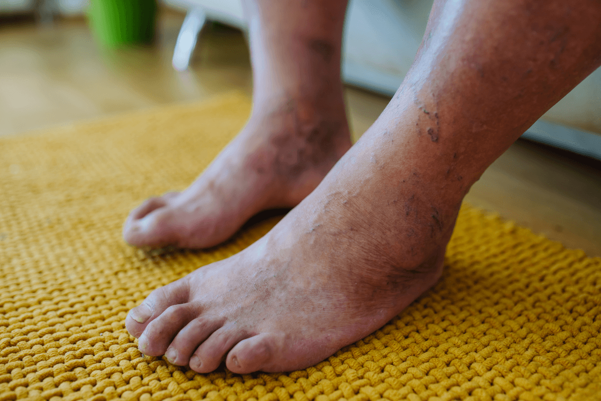
    Common diabetic foot ulcer symptoms include swelling, warmth, drainage on socks, and skin that looks white or soggy around a wound. A new odor, yellow fluid, or increasing tenderness can suggest infection or tissue change. If you have neuropathy, pain may be absent even when the wound is serious.

    Online searches for diabetic feet pictures beginning stages or early stage foot ulcer pictures can give a rough idea of what wounds may look like. Still, photos can mislead. Lighting, skin tone, camera angle, and wound depth all change what you see. A clinician can examine the foot, check circulation, and decide whether deeper tissue is involved.

    Quick tip: Take photos in the same lighting and distance if you are tracking changes between appointments.

    Call a clinician soon Seek urgent care today
    • New open area
    • Drainage or wet dressing
    • Increasing edge redness
    • Blister after shoe rubbing
    • Fever or chills
    • Rapidly spreading redness
    • Black, gray, or pale tissue
    • Severe pain or sudden color change

    If you feel unwell, or the foot changes quickly, it is reasonable to seek urgent evaluation. A foot infection can progress faster in people with diabetes, especially when blood flow is reduced.

    Why Foot Ulcers Happen In Diabetes

    Most diabetic foot ulcers develop from several factors working together. Neuropathy (nerve damage) can reduce protective sensation, so rubbing, heat, pressure, or a cut may go unnoticed. Over time, repeated pressure can break down skin, especially under the ball of the foot, heel, or toe joints.

    Blood flow also matters. Peripheral artery disease (narrowed leg arteries) can limit oxygen delivery to the skin. When circulation is reduced, even a small wound may close slowly. Smoking, kidney disease, long-standing diabetes, and previous ulcers can increase risk.

    Foot shape and footwear are often part of the story. Hammertoes, bunions, high arches, or a prominent bone can create pressure points. Shoes that once fit well may become risky if swelling, deformity, or reduced sensation changes how the foot bears weight.

    Nerve changes are important because they change how the foot reports danger. For deeper background on numbness, burning, and sensation loss, read Diabetic Neuropathy.

    High glucose is not the only cause, but it can affect inflammation and immune response. If you are noticing more thirst, frequent urination, or repeated high readings, Symptoms Of Hyperglycemia reviews common signs to discuss with a clinician.

    How Clinicians Assess A Foot Wound

    A good assessment looks beyond the visible sore. Clinicians usually measure wound length, width, and depth. They check the edges, drainage, surrounding skin, odor, and whether dead tissue is present. They also look for pressure points that may keep the wound open.

    Circulation testing is common. This may include checking pulses, skin temperature, capillary refill, and tests such as an ankle-brachial index, often called an ABI. If blood flow is limited, a vascular referral may be needed. Imaging, such as an X-ray, can be used when bone infection or a foreign object is a concern.

    Nerve testing may use a monofilament, which is a soft filament used to check protective sensation. Loss of protective sensation helps explain why diabetic foot ulcers can grow without much pain. Foot shape, gait, toe alignment, and shoe wear patterns also help the care team find the source of pressure.

    Because diabetes affects the whole body, routine care often includes broader monitoring. People reviewing age-related diabetes planning may find Diabetes Management In Older Adults useful for caregiver discussions and daily routines.

    Treatment Basics: Cleaning, Dressings, And Offloading

    Diabetic foot ulcer treatment usually focuses on three goals: protect the wound, reduce pressure, and treat infection or circulation problems when present. The exact plan depends on wound depth, location, drainage, blood flow, and your overall health history.

    Many care plans include gentle cleaning and a dressing that supports moisture balance. Some wounds need more drainage control. Others need protection from drying out. A clinician may remove dead or thickened tissue, a process called debridement, when it is appropriate. This can help reveal the true wound size and reduce barriers to healing.

    Offloading is often central. This means taking pressure off the wound with a removable boot, special shoe, insert, felt padding, or a cast in selected cases. A dressing cannot do its job if the same spot is stressed with every step. Even short barefoot walks at home can reopen fragile tissue.

    What To Ask About Your Dressing Plan

    Dressing instructions should be specific. Ask how often to change it, how to cleanse the area, what drainage is expected, and which changes should trigger a call. Avoid soaking the foot or using harsh antiseptics unless your clinician recommends them. If reaching the foot is difficult, a family member, caregiver, or home nursing support may make the routine safer.

    People often search for the best ointment for diabetic foot ulcer or the best antibiotic cream for diabetic foot ulcer. There is no single product that fits every wound. Ointments, creams, and dressings depend on infection signs, moisture needs, skin condition, and wound depth. Using the wrong product can irritate fragile skin or trap too much moisture.

    If skin issues overlap with rashes, cracks, or fungal irritation, the Dermatology category may help you browse related skin-care topics. Use it for orientation, not as a substitute for wound assessment.

    Infection, Antibiotics, And Safety Questions

    Antibiotics are not used for every open wound. Diabetic foot ulcer treatment antibiotic decisions usually depend on signs of infected tissue, not the presence of a sore alone. Clinicians look for warmth, swelling, increasing pain, pus, spreading redness, fever, or feeling generally unwell.

    When infection is suspected, the wound may be cleaned or debrided before a culture is taken. This can help identify likely bacteria and guide medication choices. Treatment decisions also consider kidney function, allergies, other medicines, and whether the infection may involve deeper tissue or bone.

    Some readers ask, can you die from diabetic foot ulcer. A foot ulcer itself is not usually the direct cause of death, but complications can become life-threatening. Severe infection, sepsis, poor circulation, kidney disease, heart disease, and complications after major surgery can all raise risk. This is why early wound care, infection control, and circulation assessment matter.

    Questions such as why do diabetics die after amputations or how long do diabetics live after amputations are understandable, but the answers vary widely. Amputation often happens in people who already have advanced infection, poor blood flow, or other serious health problems. Survival depends on age, heart and kidney health, circulation, infection control, mobility, and follow-up care. A care team can explain the individual risks more accurately than a general statistic.

    Why it matters: A worsening wound is a medical signal, not just a skin problem.

    Tracking Healing And Reducing Repeat Wounds

    Healing is usually tracked by wound size, depth, drainage, odor, surrounding redness, and the quality of new tissue. A wound that becomes smaller, has less drainage, and develops healthier-looking edges is often moving in the right direction. A wound that stops improving, smells different, or becomes more red needs reassessment.

    Some people ask whether diabetic foot ulcers can be cured. Many ulcers can close with appropriate care, pressure relief, infection management, and circulation support. However, a healed ulcer does not mean the risk is gone. The skin remains vulnerable, and the pressure pattern that caused the wound may still exist.

    Prevention focuses on daily inspection, protective footwear, careful nail care, and follow-up. Moisturize dry skin, but avoid putting lotion between toes where dampness can build. Shake out shoes before wearing them. Check for pebbles, rough seams, or worn insoles. Bring your shoes to appointments so clinicians can identify pressure areas.

    Glucose tracking can also help your team see patterns that may affect healing. Some people use meters, continuous glucose monitors, or prescribed medications as part of their diabetes plan. For device context, OneTouch Verio Flex Meter and Dexcom G7 Sensor are examples of glucose monitoring options to recognize when discussing supplies with a clinician.

    Medication reviews may also be part of wound care, especially when glucose levels are frequently high or low. If you are comparing common diabetes medication names, Metformin and Lantus Insulin Vial are examples of product pages that may help you identify items in your medication list. Do not change diabetes medicines without prescriber guidance.

    For broader diabetes education, the Diabetes category organizes related topics in one place.

    Authoritative Sources

    The American Diabetes Association explains foot screening and prevention in its resource on diabetes-related foot complications.

    The Infectious Diseases Society of America provides clinical guidance on evaluating and treating diabetic foot infections.

    The NCBI Bookshelf clinical review summarizes risk factors and complications in diabetic foot ulceration.

    Recap

    Diabetic foot ulcers often start small, but they deserve early attention. Watch for blisters, cracks, drainage, calluses with dark centers, spreading redness, odor, fever, or sudden color changes. The core of care is assessment, pressure relief, wound protection, infection management when needed, and follow-up.

    If a wound changes quickly or you feel unwell, seek prompt medical evaluation. For ongoing questions, a podiatrist, wound clinic, or diabetes care team can tailor advice to your health history, circulation, footwear, and medication plan.

    This content is for informational purposes only and is not a substitute for professional medical advice.

  • Using HEMANGEOL for Infantile Hemangiomas Safely

    Using HEMANGEOL for Infantile Hemangiomas Safely

    HEMANGEOL is an oral propranolol treatment used for some infantile hemangiomas that need medicine rather than watchful waiting. The core of Hemangiol Medicine for Infantile Hemangiomas: Safety Tips is simple: safe use depends on regular feeding, careful monitoring, and fast action if a baby seems unusually sleepy, wheezy, pale, weak, or hard to wake. This matters because many hemangiomas never need systemic treatment, while the ones that do may threaten vision, breathing, feeding, skin integrity, or later appearance. Parents and caregivers usually need a calm safety plan, not vague reassurance.

    Key Takeaways

    • HEMANGEOL is a liquid beta blocker used when some infantile hemangiomas need whole-body treatment.
    • Low blood sugar is a major concern, especially during poor feeding, vomiting, or illness.
    • Breathing problems, slow heart rate, low blood pressure, and unusual sleepiness need prompt attention.
    • Many infantile hemangiomas can be watched; treatment is usually reserved for higher-risk cases.
    • Ask early about feeding rules, monitoring, sick-day instructions, and when to seek urgent care.

    Hemangiol Medicine for Infantile Hemangiomas: Safety Tips That Matter

    HEMANGEOL is a liquid form of propranolol, a beta blocker (a medicine that blocks certain stress signals in the body). It is used for infantile hemangioma when a clinician believes systemic treatment (medicine that works throughout the body) is needed. At a high level, propranolol appears to slow blood-vessel growth signals and reduce blood flow within the lesion, which can help limit growth and improve how the area looks over time.

    Not every infantile hemangioma needs treatment. Many grow for a period, then slowly flatten and fade without medicine. Treatment is more likely to be considered when the spot is ulcerating, painful, bleeding, growing quickly, or located where it can affect vision, breathing, feeding, hearing, or the shape of the nose, lip, or other facial structures. Example: a small superficial mark on the trunk may be watched, while a fast-growing lesion near an eyelid raises a different level of concern.

    Why it matters: Safety questions only make sense when the reason for treatment is clear.

    Timing matters too. Infantile hemangiomas often change fastest in early infancy, so the decision to start or defer treatment can look different from one visit to the next. A lesion that seems low risk at first may become more concerning if it blocks a natural opening, breaks down, or starts to distort nearby tissue. That is why the goal is not to treat every hemangioma, but to identify the ones where early action may protect function or reduce lasting change.

    That is also why parents are often weighing watchful waiting against active treatment, not simply choosing a medicine. For broader child-health and skin-condition reading, the site’s Pediatrics hub and Dermatology hub offer related background. A specialist may also look at how deep the hemangioma is, whether it is still in a rapid-growth phase, and how likely it is to leave lasting distortion if nothing is done.

    BorderFreeHealth works with licensed Canadian partner pharmacies for cross-border prescription access.

    Main Risks, Side Effects, and Red Flags

    The most important HEMANGEOL safety issues are low blood sugar, breathing problems, and effects on heart rate or blood pressure. HEMANGEOL side effects in babies can also include sleep changes, cooler hands or feet, fussiness, and stomach upset. Those milder effects are not always dangerous, but they still matter because they can be mistaken for normal infant behavior.

    Low blood sugar deserves special attention. Beta blockers can make some warning signs harder to notice, and the risk can rise when a baby is feeding poorly, vomiting, or going longer than usual without calories. Caregivers may be told to watch for unusual sleepiness, limpness, shakiness, sweating, pallor, poor feeding, or, in severe situations, a seizure. That is one reason feeding instructions are so central to safe treatment.

    Breathing and circulation issues also need respect. Propranolol may worsen wheezing in susceptible babies, and it can lower heart rate or blood pressure. A baby who is struggling to breathe, turning blue, collapsing, having a seizure, or difficult to wake needs urgent evaluation. Less dramatic changes, such as new wheezing, repeated vomiting, marked weakness, or persistent poor intake, still deserve prompt contact with the care team.

    Concern What caregivers may notice Why clinicians ask about it
    Low blood sugar Sleepiness, pallor, shakiness, poor feeding, limpness Risk rises when feeds drop or illness interrupts intake
    Breathing problems Wheezing, cough with breathing difficulty, working hard to breathe Beta blockers can aggravate bronchospasm in some infants
    Slow heart rate or low blood pressure Unusual weakness, faint-looking episodes, poor responsiveness Monitoring helps detect tolerance problems early
    Sleep or behavior changes Restless sleep, more night waking, unusual irritability Common worries are often manageable but worth discussing

    Another practical point is that not every side effect points to danger, but repeated patterns matter. If feeds get shorter, naps change abruptly, or the baby seems less interactive after doses, that trend is worth documenting and reporting. A simple notebook or phone note can keep one odd day from being confused with a true medication pattern.

    Sleep problems deserve a separate mention because they are common online search questions. Some babies become more wakeful or restless, while others seem sleepier. Most of these reports are not emergencies. Still, a baby who is far harder to rouse than usual should not be written off as simply having a bad nap day. The difference between a nuisance effect and a red flag is how alert, pink, and responsive the child is overall.

    Feeding, Illness, and Monitoring Are Central

    Feeding and monitoring are central because low blood sugar risk rises when a baby is not taking normal feeds. Caregivers starting HEMANGEOL usually need very practical answers: when the medicine should be given in relation to feeding, what to do during vomiting or poor intake, and which symptoms mean the next dose plan needs to be reviewed.

    Before or around the start of treatment, clinicians often review weight, feeding pattern, birth history, heart or lung concerns, and any other medicines. Some teams also check heart rate and blood pressure at the start or after changes, depending on the baby’s age, health status, and treatment setting. If a baby may need to fast for a test or procedure, that question should be settled ahead of time rather than on the day.

    Questions to have answered early

    • Why treatment now — what specific risk is being prevented?
    • How should doses relate to feeds or bottles?
    • What should happen if vomiting starts or intake drops?
    • Which symptoms need a same-day call?
    • Which signs mean urgent care is needed?
    • How will follow-up and monitoring be handled?
    • Are any other medicines or conditions relevant?

    Quick tip: Keep one written plan for feeds, doses, and red-flag symptoms.

    It also helps to use the measuring device supplied by the pharmacy and to store the bottle safely out of reach. A written medication guide is worth keeping close, especially for anyone else who may feed the baby or give a dose. Grandparents, babysitters, and daycare staff do not need a full lecture, but they do need the same key safety rules.

    A monitoring plan should also cover who to call after hours and how to mention the medicine if another clinician sees the child for an unrelated illness. Urgent care or emergency staff may not know the hemangioma history, but they still need to know the baby is taking a beta blocker. That small detail can shape safer decision-making during a stressful visit.

    When required, a pharmacy checks prescription details with the original prescriber before dispensing.

    Who Needs Extra Caution Before Starting

    Extra caution matters when a baby has other health issues that could raise the risk of beta-blocker side effects. The care team may ask about wheezing, bronchiolitis, breathing pauses, heart disease, rhythm problems, low blood sugar history, poor feeding, low weight, or any condition that makes intake unpredictable. This is not about excluding every child from treatment. It is about deciding what level of monitoring makes sense.

    Medication review matters too. Families should tell the prescriber about every prescription drug, over-the-counter product, or supplement the baby is receiving, because even an unrelated medicine can change how safely a beta blocker fits into care. Illness matters just as much. A baby with diarrhea, repeated vomiting, or a respiratory infection may need a specific sick-day plan rather than a guess made in the moment.

    Premature infants or babies with complex medical histories may not follow the standard script. That does not automatically rule out treatment, but it can make coordination between pediatrics, dermatology, and sometimes cardiology more important. In that setting, the best safety plan is usually the clearest one, with each caregiver knowing the same instructions.

    Parents often ask about long-term effects. In real-world use, the immediate safety issues and the hemangioma’s response usually drive the discussion more than a single long-term internet question. Follow-up is typically shaped around growth of the lesion, tolerance of the medicine, and the child’s overall health. If you are unsure why therapy is continuing or being changed, that is a strong reason to ask for the treatment goal in plain language.

    How It Compares With Other Treatment Paths

    HEMANGEOL and generic propranolol share the same active ingredient, but they are not casual substitutes. Families often search HEMANGEOL vs propranolol as if they were different drugs. The better question is whether the exact formulation, concentration, labeling, measuring device, and instructions match the prescription. That is especially important with liquid medicines, where a mix-up can create avoidable risk.

    Comparison also matters at the treatment-plan level. Some infantile hemangiomas can be observed without medicine. Some smaller or more superficial lesions may be considered for topical beta-blocker treatment in selected cases. Others, especially those threatening function or causing ulceration, may need whole-body therapy or closer specialist input. The choice depends on location, depth, growth behavior, and complication risk, not just on how dramatic the lesion looks in a photo.

    Families sometimes focus on before-and-after photos. Those images can be reassuring, but they do not show why one baby was treated while another was watched, or how much monitoring happened behind the scenes. A safer comparison looks at function, growth risk, and tolerance, not just appearance.

    If you are trying to make sense of options, ask what problem the treatment is supposed to solve: stopping rapid growth, preventing skin breakdown, protecting vision or breathing, or reducing the chance of lasting distortion. That question usually brings more clarity than asking which medicine is strongest. For browseable site sections rather than treatment advice, the Dermatology Products hub can help you explore related skin-care topics.

    Some eligible patients without insurance explore cash-pay cross-border prescription options.

    Authoritative Sources

    Further reading can help you ask better questions, but the safest plan is the written one from your child’s prescriber and pharmacy. In most cases, the big safety themes are clear: understand why treatment is needed, link dosing to feeding exactly as directed, know the red flags, and do not guess during illness or poor intake. That frame can make a confusing topic feel much more manageable.

    This content is for informational purposes only and is not a substitute for professional medical advice.
  • Hair Loss in Young Adults: Why It’s Rising and Next Steps

    Hair Loss in Young Adults: Why It’s Rising and Next Steps

    Hair Loss in Young Adults: Why It’s Rising and Next Steps starts with a simple answer: early thinning is usually driven by overlapping factors, not one hidden mistake. Genetics, hormones, stress, nutrition, scalp inflammation, tight hairstyles, illness, and some medical conditions can all contribute. Why this matters: the right next step depends on the pattern, speed, symptoms, and health history. A receding hairline, sudden shedding, round patches, and scaling scalp changes point to different possibilities.

    Key Takeaways

    • Early thinning has many causes, including genetics, stress, nutrition, hormones, infections, and styling tension.
    • Sudden shedding differs from gradual pattern loss, so timing and scalp symptoms matter.
    • A clinician may review your history, scalp, medications, nutrition, and blood tests before suggesting care.
    • Treatment works best when it targets the cause, not the trend or internet diagnosis.
    • Seek care sooner for patchy loss, pain, scaling, scarring, rapid changes, or other new symptoms.

    Why Hair Loss Is Rising in Young Adults

    Early hair concerns seem more visible because young adults face more overlapping pressures and notice changes sooner. Social media, frequent photos, and close-up video calls can make normal variation feel alarming. At the same time, real contributors may be increasing for some people. These include chronic stress, disrupted sleep, restrictive eating, rapid weight changes, intense styling, scalp irritation, and delayed care for medical symptoms.

    This does not mean every young person with thinning has permanent baldness. Some shedding is temporary, especially after illness, major stress, surgery, childbirth, or a significant diet change. Other patterns, such as androgenetic alopecia, can progress gradually without early support. The goal is to separate short-term shedding from ongoing follicle miniaturization, inflammation, infection, or traction damage.

    Why it matters: Early assessment can prevent months of guessing and unnecessary products.

    Young people may also search for answers earlier than past generations. That can make the issue look like a sudden epidemic. Still, the concern is valid. Hair changes can affect confidence, identity, dating, school, work, and mental health. Taking the concern seriously is not vanity. It is part of whole-person care.

    Shedding, Thinning, and Pattern Loss Are Not the Same

    The first useful distinction is whether you are shedding more hairs or losing visible density over time. Shedding means more hairs are falling out than usual. Thinning means the scalp looks more visible, the ponytail feels smaller, or the hairline changes. Both can happen together, but they can come from different causes.

    Hair grows in cycles. A trigger can push more hairs into the resting phase, leading to telogen effluvium, a form of temporary shedding. Pattern loss usually involves gradual miniaturization, where hairs grow finer over time. Patchy loss can point toward alopecia areata, an autoimmune condition where the immune system affects hair follicles. Tight styles can cause traction alopecia, which comes from repeated pulling on the follicle.

    What You Notice Common Pattern Possible Clues
    More hair in the shower or brush Increased shedding Recent stress, illness, diet change, childbirth, or medication review
    Temple recession or crown thinning Pattern loss Family history, gradual change, finer hairs in affected areas
    Round or sharply defined patches Patchy loss Smooth patches, nail changes, autoimmune history, sudden onset
    Breakage near tight styles Traction or breakage Braids, extensions, tight ponytails, pain, bumps, or edge thinning

    Hair shedding vs hair loss can feel hard to judge at home. Photos taken in the same light can help show whether density is changing. A drain full of hair after washing is not enough by itself, especially if you wash less often. The broader pattern matters more than one alarming shower.

    Common Causes Young Adults Should Discuss

    Young adult hair loss causes often overlap, so a single label may not explain everything. A person can have inherited pattern thinning and also experience temporary shedding after stress or illness. Another person may have iron deficiency, thyroid disease, scalp inflammation, or traction damage. This is why a careful history matters.

    Genetic and hormonal patterns

    Androgenetic alopecia in young adults is common and can affect people of any sex. It often runs in families, but family history is not always obvious. In men, it may show as a receding hairline in your 20s, temple recession, or crown thinning. In women, it may show as widening of the part, reduced volume, or diffuse thinning over the top of the scalp.

    Hormonal changes can also influence the hair cycle. Conditions that affect androgens, thyroid hormones, or reproductive hormones may contribute to thinning or shedding. Clues can include acne, irregular periods, new facial hair growth, fatigue, weight changes, heat or cold intolerance, or changes in mood and energy. These signs deserve medical discussion rather than self-treatment.

    Stress, illness, and nutrition

    Stress related hair loss often means the body has shifted more hairs into a resting phase. This can happen after intense emotional stress, high fever, major illness, surgery, childbirth, or rapid weight change. The shedding may appear after the trigger, which can make the connection easy to miss.

    Nutritional deficiency hair loss can occur when the body lacks key building blocks. Iron deficiency is a common concern, especially in people with heavy menstrual bleeding, restrictive diets, low intake of iron-rich foods, or digestive conditions that affect absorption. Vitamin deficiency hair loss is possible, but supplements are not automatically the answer. Too much of some nutrients can be harmful, and not every deficiency causes thinning.

    Protein intake, overall calories, and eating patterns matter too. Young adults may experiment with intense diets, fasting, or high-pressure fitness routines. A clinician or registered dietitian can help identify whether nutrition is a likely contributor without turning food into another source of stress.

    Scalp, immune, and styling factors

    Alopecia areata in young adults can cause round patches or sudden loss in specific areas. It is not caused by poor hygiene or a bad routine. Scalp infections can also cause shedding, scaling, itching, tenderness, or broken hairs. These need proper diagnosis because cosmetic products cannot treat an infection.

    Traction alopecia from hairstyles deserves special attention. Tight braids, extensions, buns, ponytails, and repeated edge tension can damage follicles over time. Early traction changes may improve when tension stops. Long-standing traction may become harder to reverse, especially if scarring develops.

    Hair practices can also cause breakage without true follicle loss. Bleaching, heat styling, chemical straightening, and harsh brushing can weaken the hair shaft. Breakage often leaves uneven short hairs rather than full strands with bulbs at the end.

    What a Hair Loss Evaluation Usually Includes

    A good evaluation starts with your story, not a product recommendation. A clinician may ask when the change began, how fast it progressed, whether shedding is diffuse or patchy, and whether the scalp itches, hurts, flakes, or burns. They may also ask about family history, menstrual changes, pregnancy, recent illness, stress, diet, weight changes, medications, supplements, and hair practices.

    The scalp exam can reveal important clues. Redness, scaling, pustules, scarring, broken hairs, miniaturized hairs, or sharply defined patches can guide the next step. Some clinicians may perform a gentle pull test, examine the scalp closely, or order blood tests. Common lab discussions may include iron status, thyroid function, and other tests based on symptoms and history.

    Example: a student notices sudden shedding after a severe illness and a stressful exam period. Another person notices a slowly widening part and a strong family history of early thinning. These examples can look similar in the shower, but they may need different evaluations.

    Seek care sooner if hair loss is sudden, patchy, painful, associated with scaling or sores, or accompanied by fatigue, weight change, fever, irregular periods, or other new symptoms. A dermatologist can be especially helpful when the diagnosis is unclear or the scalp looks inflamed. For broader skin and hair topics, you can browse Dermatology Topics for related educational reading.

    Treatment Options Depend on the Cause

    The most helpful treatment plan matches the cause and the person’s goals. Temporary shedding may improve as the trigger resolves, but a clinician may still check for deficiencies or medical conditions. Pattern thinning may involve topical options, prescription medicines for selected adults, procedures, or camouflage strategies. Patchy autoimmune loss, infections, inflammatory scalp disease, and traction damage need different approaches.

    Topical minoxidil is one commonly discussed option for pattern thinning, but it is not the right answer for every situation. Age, pregnancy plans, scalp irritation, other medicines, and diagnosis all matter. Oral prescription options may be considered for some adults, but they have important safety considerations and are not appropriate for everyone. Do not start, stop, or combine treatments without medical guidance.

    For scalp infections, antifungal or other prescription treatment may be needed. For alopecia areata, clinicians may discuss anti-inflammatory treatments or other options depending on the extent and location. For traction alopecia, reducing tension is central. This may mean loosening styles, taking breaks from extensions, avoiding painful hairstyles, and treating scalp inflammation early.

    BorderFreeHealth works with licensed Canadian partner pharmacies for eligible U.S. patients.

    If a prescribed dermatology medicine becomes part of your care plan, compare only after the diagnosis and prescription are clear. The Dermatology Products hub is a browsable product category, not a substitute for medical evaluation. For some people without insurance, cash-pay access conversations may be relevant, subject to eligibility and local rules.

    A Practical Next-Step Checklist

    You do not need a perfect answer before asking for help. Bring clear observations and avoid changing too many things at once. That makes it easier to see what is helping and what is not.

    • Take baseline photos in consistent lighting.
    • Note when shedding or thinning started.
    • List recent illness, stress, weight, or diet changes.
    • Bring medication and supplement names.
    • Describe itching, pain, scaling, or redness.
    • Review tight styles, extensions, heat, and chemicals.
    • Ask which labs are reasonable for your symptoms.
    • Discuss treatment goals and safety concerns.

    Prescription details may be checked with the prescriber before pharmacy dispensing when required.

    Avoid shaving your head, starting several supplements, changing all hair products, and beginning multiple treatments on the same day unless a clinician recommends it. When everything changes at once, it becomes harder to know what caused improvement or irritation.

    How to Protect Hair and Scalp Health Day to Day

    Prevention is not about controlling every strand. It is about reducing avoidable stress on the hair cycle and scalp. Gentle hair handling, adequate nutrition, sleep, and prompt care for scalp symptoms can support healthier growth conditions. These steps cannot override genetics, but they can reduce preventable breakage and traction.

    Use styles that do not hurt. Pain, bumps, or headaches from a hairstyle are warning signs. Rotate styles when possible, avoid constant tension on the same area, and be cautious with heavy extensions. If you chemically treat or bleach your hair, spacing services and using heat carefully may reduce breakage.

    Nutrition should be steady and realistic. Hair is not the body’s top survival priority, so rapid dieting, low protein intake, or untreated deficiencies can show up in shedding. Still, more supplements do not mean more growth. Testing and targeted correction are safer than stacking vitamins based on social media advice.

    Sleep and stress support also matter, even when they do not fully explain the problem. Stress can worsen shedding, scalp picking, and inflammation. If hair changes are increasing anxiety, support from a clinician, therapist, or trusted community can be part of care.

    The Emotional Side of Early Thinning

    Hair changes in your teens or 20s can feel isolating. Friends may dismiss it, while online searches can make every possibility sound urgent. You may feel pressure to hide, over-style, or spend money on products before you know the cause. Those reactions are understandable.

    Try to separate appearance concerns from blame. Hair loss is not a character flaw, and it is not proof that you failed at health. A calm plan can protect both your scalp and your confidence. If you remember one point from Hair Loss in Young Adults: Why It’s Rising and Next Steps, make it this: pattern, timing, and symptoms should guide decisions.

    Cash-pay cross-border options can depend on eligibility and jurisdiction.

    Authoritative Sources

    Further Reading and Recap

    Early thinning can come from inherited patterns, temporary shedding, deficiencies, thyroid or hormonal changes, autoimmune conditions, infections, traction, or breakage. The safest next step is to document the pattern, check for symptoms, and seek an evaluation when changes are persistent, sudden, patchy, painful, or distressing.

    With the right diagnosis, care can become more focused and less reactive. You do not need to solve it alone, and you do not need to chase every trend before getting a clear assessment.

    This content is for informational purposes only and is not a substitute for professional medical advice.
  • Metformin and Hair Loss: Evidence, CCCA, and Recovery

    Metformin and Hair Loss: Evidence, CCCA, and Recovery

    Metformin and hair loss are not strongly linked by current evidence, but hair changes can still happen around the same time for other reasons. Metformin may indirectly affect shedding through vitamin B12 deficiency, digestive side effects, weight or diet changes, PCOS-related hormone shifts, or the underlying condition being treated. In central centrifugal cicatricial alopecia (CCCA), the stakes are different because inflammation can permanently damage follicles.

    Why this matters: not all thinning has the same cause. Temporary shedding, androgen-related thinning, and scarring alopecia need different levels of urgency and different conversations with your clinician.

    Key Takeaways

    • Direct link unclear: metformin is not widely recognized as a common hair-loss cause.
    • B12 matters: long-term use can lower vitamin B12 in some people.
    • CCCA is urgent: scarring hair loss needs early dermatology care.
    • PCOS can confuse timing: hormones and insulin resistance also affect hair.
    • Tracking helps: photos, symptoms, labs, and medication timing guide evaluation.

    Does Metformin Cause Hair Loss or Thinning?

    For most people, metformin is not considered a typical direct cause of hair loss. Reports of shedding after starting treatment do occur, but timing alone does not prove causation. Hair growth cycles respond slowly to illness, stress, nutrition changes, hormone shifts, inflammation, and medications, so the real trigger may have started weeks earlier.

    The most common metformin side effects are gastrointestinal, such as nausea, diarrhea, abdominal discomfort, and appetite changes. Those effects can indirectly affect nutrition in some people. If eating patterns change sharply, or if weight loss is rapid, the body may shift more hairs into the shedding phase. Dermatologists call this telogen effluvium, a non-scarring shed where follicles usually remain alive.

    Metformin and hair loss can also overlap because of the conditions metformin treats. Type 2 diabetes, insulin resistance, PCOS, thyroid disease, iron deficiency, and chronic inflammation can all influence hair density. That is why a medication review should sit beside a broader hair and scalp evaluation, not replace it.

    If you want context on why clinicians use this medicine in metabolic care, this overview of Metformin Benefits explains its wider role without focusing only on hair.

    Why CCCA Changes the Conversation

    CCCA is a scarring alopecia, meaning inflammation can replace active follicles with scar tissue. It often starts around the crown and may cause tenderness, itching, burning, breakage, or gradual thinning. It is more common in women of African descent, although individual risk varies.

    In CCCA, the main question is not only whether a medication caused shedding. The more urgent question is whether active scalp inflammation is damaging follicles right now. Once a follicle is scarred, regrowth is limited. Early diagnosis and inflammation control can help protect remaining hair.

    Some emerging research has explored metformin in scarring alopecia and fibrosis pathways, including topical or local approaches in specialist settings. That does not mean oral metformin should be started, stopped, or adjusted for hair loss without medical review. Evidence is still developing, and CCCA care usually involves a dermatologist-led plan.

    For broader hair-care context and treatment categories, see Hair Loss Treatment. If you want to browse related scalp and skin topics, the Dermatology section can help you compare nearby conditions.

    Quick tip: New crown tenderness, burning, scaling, or rapid widening deserves prompt scalp evaluation.

    B12 Deficiency, Shedding, and Lab Checks

    Vitamin B12 deficiency is one of the clearest indirect issues to consider with long-term metformin use. Metformin can reduce B12 absorption in some people, especially with longer use or other risk factors. Low B12 can contribute to anemia, fatigue, numbness or tingling, mouth soreness, and neurologic symptoms.

    B12 deficiency is not the only nutritional cause of hair shedding. Ferritin, which reflects iron stores, may also matter. Thyroid imbalance, low vitamin D in some contexts, restrictive diets, recent illness, and major stress can all add to shedding risk. The practical point is simple: avoid guessing from timing alone.

    Ask your clinician whether labs fit your situation. Common discussions include vitamin B12, complete blood count, ferritin, thyroid-stimulating hormone (TSH), and sometimes androgen testing when PCOS or patterned thinning is suspected. These tests do not diagnose every hair disorder, but they can remove common confounders.

    For product-level medication context, the Metformin page can help you identify the medicine name before discussing it with a clinician. BorderFreeHealth connects U.S. patients with licensed Canadian partner pharmacies, and prescription details are verified when required before pharmacy dispensing.

    PCOS, Androgens, and Women’s Hair Patterns

    Women asking whether metformin causes hair loss often have another overlapping factor: PCOS. Polycystic ovary syndrome can involve insulin resistance, irregular cycles, acne, excess facial or body hair, and higher androgen activity. Androgens can miniaturize scalp follicles in a patterned way, often around the part line or crown.

    Metformin may be used in PCOS care to support insulin sensitivity, but its effects on scalp hair are not simple or guaranteed. Some people notice less hormone-related worsening when metabolic factors improve. Others continue to have thinning because genetics, androgen sensitivity, iron status, thyroid function, or CCCA are also involved.

    Hirsutism, which means excess coarse hair growth in androgen-sensitive areas, is different from scalp shedding. A person can have more facial hair and less scalp density at the same time. That pattern points clinicians toward hormone evaluation rather than a single medication explanation.

    Related hair patterns can overlap. For example, Male Pattern Baldness explains androgen-driven miniaturization in a different population, but the follicle biology helps show why diagnosis matters.

    Recovery: What to Expect Before Changing Anything

    Metformin hair loss recovery depends on what is actually causing the change. If the issue is telogen effluvium from illness, stress, rapid dietary change, or a temporary metabolic shift, shedding may improve after the trigger is addressed. If low B12 or iron is contributing, correction may help reduce ongoing shedding, though visible density changes usually take time.

    If CCCA is active, recovery has a different meaning. The goal is often to reduce symptoms, slow progression, and protect remaining follicles. Regrowth may be limited in scarred areas, which is why early treatment discussions are important. A scalp biopsy may be considered when the diagnosis is uncertain.

    Do not stop metformin on your own because hair shedding begins. Stopping or changing diabetes or PCOS treatment can affect glucose control and other health goals. Instead, bring a clear timeline to the clinician who prescribed it and, when possible, to a dermatologist familiar with hair loss.

    Example Timeline to Bring to a Visit

    • Medication start: note date, dose changes, and formulation changes.
    • Shedding onset: record when you first noticed extra hair fall.
    • Scalp symptoms: include pain, itch, burning, flakes, or pustules.
    • Health events: list illness, surgery, stress, or rapid weight change.
    • Hair practices: note traction styles, relaxers, heat, or chemical processing.

    Monthly photos can help. Use the same room, lighting, hairstyle, and angles. Photograph the crown, part line, temples, and any tender areas. These images can show whether the pattern is diffuse shedding, patterned thinning, breakage, or a scarring pattern.

    Medication Formulation, Dose Timing, and Other Clues

    Questions about metformin dosage and hair loss usually need careful interpretation. A person may notice shedding after starting metformin 500 mg, after increasing the dose, or after switching formulations. That pattern is worth documenting, but it still does not prove the medicine directly caused follicle damage.

    Digestive intolerance can be a clue. If nausea or diarrhea leads to poor intake, fewer protein-rich meals, or missed supplements, nutrition may become part of the problem. Some clinicians consider formulation changes when gastrointestinal effects persist, but that decision belongs in medical care, not trial-and-error self-management.

    People also ask about brand and generic names. Glucophage is a brand name for metformin, while metformin is the generic drug name. For a neutral comparison of naming and formulation context, see Glucophage vs Metformin.

    Practical Questions to Ask Your Clinician

    A focused appointment works better than a broad worry about hair loss. Bring your medication list, photos, timeline, and scalp symptoms. If CCCA is possible, ask whether you need referral to a dermatologist who evaluates scarring alopecia.

    • Diagnosis question: is this shedding, breakage, patterned thinning, or scarring alopecia?
    • Lab question: should B12, ferritin, CBC, thyroid, or androgens be checked?
    • Medication question: could side effects be affecting nutrition or adherence?
    • Scalp question: are inflammation, scale, tenderness, or pustules present?
    • Care question: which hair practices should be modified to reduce traction?

    Some people explore topical hair products while awaiting evaluation. Options vary by diagnosis, and not every product fits scarring alopecia. If a clinician recommends reviewing topical categories, Minoxidil and Minoxidil Foam are product references you can use for label and ingredient discussions. For inflammatory scalp conditions, Clobetasol may also appear in treatment conversations, but steroid use should be clinician-directed.

    Why it matters: The wrong assumption can delay care for a scarring condition.

    Authoritative Sources

    The FDA label for metformin tablets lists established prescribing information, contraindications, and safety details, including vitamin B12 monitoring language.

    The American Academy of Dermatology provides a patient resource on central centrifugal cicatricial alopecia, including signs, risk context, and why early care matters.

    The American Diabetes Association publishes Standards of Care in Diabetes, which discuss medication monitoring considerations, including periodic B12 assessment in long-term metformin use.

    Recap

    Metformin and hair loss should be evaluated with nuance. Current evidence does not show metformin as a common direct cause of hair loss, but B12 deficiency, digestive side effects, PCOS, diabetes, thyroid disease, iron status, and stress can all shape hair changes. In CCCA, prompt scalp-focused care is especially important because scarring can be permanent.

    Your next best step is not to guess from online stories. Build a timeline, check for scalp symptoms, ask about relevant labs, and seek dermatology input when crown thinning, tenderness, burning, or scarring alopecia is possible.

    This content is for informational purposes only and is not a substitute for professional medical advice.

  • Palmoplantar Pustulosis vs Dyshidrotic Eczema: Key Differences

    Palmoplantar Pustulosis vs Dyshidrotic Eczema: Key Differences

    Palmoplantar pustulosis vs dyshidrotic eczema usually comes down to what the bumps contain, how the surrounding skin looks, and what pattern repeats over time. Palmoplantar pustulosis, often shortened to PPP, tends to cause recurrent sterile pustules on red, scaly palms or soles. Dyshidrotic eczema, also called pompholyx, more often causes intensely itchy, deep-seated clear blisters that later peel. Both are noncontagious, and both can affect walking, gripping, handwashing, and sleep.

    That distinction matters because the likely triggers, helpful tests, and long-term treatment plan can differ. PPP is often discussed within the psoriasis spectrum, while dyshidrotic eczema often reflects skin-barrier irritation, allergy, sweat, or atopic tendency. A fresh flare is usually easier to interpret than dry, peeling skin several days later.

    Key Takeaways

    • PPP clue: recurrent cloudy or yellowish sterile pustules on inflamed, scaly palms or soles.
    • Eczema clue: tiny deep clear blisters, often with marked itch, followed by peeling.
    • Shared feature: both conditions are noncontagious and can flare repeatedly.
    • Trigger pattern: smoking and psoriasis clues may point toward PPP; irritants, allergens, heat, sweat, and wet work may point toward eczema.
    • Next step: photos from day one of a flare can help a clinician decide whether testing is needed.

    How Palmoplantar Pustulosis vs Dyshidrotic Eczema Looks Early

    The simplest visual difference is pustule versus vesicle. A pustule is a pus-filled bump, while a vesicle is a small fluid-filled blister. In PPP, the bumps often look cloudy, yellow, tan, or brown as they dry. In dyshidrotic eczema, the blisters usually look clear and sit deep under the skin, sometimes like tiny tapioca pearls.

    PPP often appears on a red, thickened, scaly base. The skin between flares may stay rough, cracked, or tender. The pustules are usually sterile, meaning they are not caused by a bacterial infection. Still, broken skin can become infected secondarily, so warmth, spreading redness, drainage, fever, or rapidly worsening pain deserves prompt medical review.

    Dyshidrotic eczema usually begins with sudden itch, tingling, or burning, followed by small clear blisters. These often appear along the sides of the fingers, across the palms, or on the soles. After the blister stage, the skin may peel, become dry, and crack. At that later stage, it can resemble ordinary hand or foot dermatitis.

    Why it matters: A late photo of peeling skin may hide the clue that mattered most at the start.

    Feature Palmoplantar Pustulosis Dyshidrotic Eczema
    Main bump Cloudy or yellowish sterile pustules Deep clear fluid-filled blisters
    Dominant symptom Burning, soreness, tenderness, cracking Itch is often strongest
    Background skin Red, thick, scaly, sometimes fissured May look normal early, then peel
    Common pattern Chronic relapsing pustules on palms or soles Sudden itchy flares that dry and shed
    Helpful clues Psoriasis features, nail changes, smoking association Atopy, irritants, contact allergy, heat, sweat, wet work

    A useful shorthand is clear blisters plus intense itch suggest dyshidrotic eczema, while cloudy pustules plus thick scale suggest PPP. Real skin disease is not always tidy, though. Some people have overlapping irritation, scratching, fungal infection, or contact allergy that blurs the picture.

    For broader condition education, the Dermatology collection can help you compare related skin topics.

    Symptoms That Point One Way or the Other

    The main symptom can be as helpful as the appearance. Dyshidrotic eczema is often intensely itchy, while PPP is more likely to feel sore, hot, burning, or cracked. Both can hurt when fissures open or when the soles are involved.

    Palmoplantar pustulosis symptoms

    PPP usually causes repeated crops of small pustules on the palms, soles, or both. The pustules may begin as yellow-white spots and then dry into brownish scale or crust. The surrounding skin often remains red, thickened, and scaly. Painful fissures can make walking, standing, typing, or gripping tools difficult.

    Some people also have psoriasis clues elsewhere. These may include thick plaques on the elbows, knees, scalp, or trunk, or nail changes such as pitting, lifting, or thickening. Those findings do not have to be present, but they can strengthen the psoriasis-spectrum possibility. You can learn more about related awareness and support themes in Psoriasis Action Month.

    Dyshidrotic eczema symptoms

    Dyshidrotic eczema symptoms often start with itch before the rash is obvious. The blisters tend to be small, deep, and clustered. They may appear on the sides of the fingers, palms, toes, or soles. When they dry, the skin can peel in sheets and leave tender cracks.

    Scratching can change the rash quickly. A clear blistering flare may become red, crusted, or swollen, especially if the skin barrier breaks. That is why early photos are useful. If the pattern includes repeated itchy blisters and peeling after a few days, dyshidrotic eczema becomes more likely. For a deeper look at related immune and eczema questions, see Dyshidrotic Eczema and Autoimmune Disease.

    Causes and Triggers: Why the Flare Pattern Matters

    The cause of PPP is not fully settled, but it is strongly associated with smoking and psoriasis-spectrum inflammation. Dyshidrotic eczema usually has a broader trigger profile, involving skin-barrier stress, atopy, irritants, sweating, and sometimes contact allergy.

    PPP may appear without an obvious trigger. Clinicians may still ask about tobacco exposure, psoriasis history, nail changes, recurrent infections, stress, and repeated friction on the palms or soles. Some people wonder, “How did I get palmoplantar pustulosis?” The honest answer is usually that several factors may contribute, and not every person finds a single cause.

    Dyshidrotic eczema flares often follow more everyday exposures. Wet work, frequent handwashing, harsh cleansers, occlusive gloves, sweaty shoes, heat, stress, and metal sensitivity can all play a role. Nickel and cobalt sensitivity are common contact-allergy considerations. In some cases, a fungal infection on the feet may trigger hand eczema-like flares, so clinicians may check both hands and feet.

    • Smoking exposure: more suggestive of PPP than eczema.
    • Wet work: often worsens hand eczema flares.
    • Heat and sweat: common dyshidrotic eczema triggers.
    • Thick scale: may raise psoriasis-spectrum suspicion.
    • New gloves or shoes: may suggest contact dermatitis.
    • One-sided scaling: may prompt a fungal check.

    Stress can aggravate many inflammatory skin conditions, but it rarely explains the whole picture. If stress seems tied to itchy hand flares, Stress and Dyshidrotic Eczema offers more context on that specific pattern.

    Conditions That Can Mimic Either Rash

    Several hand and foot rashes can look like palmoplantar pustulosis vs dyshidrotic eczema at first glance. This is one reason self-diagnosis from photos can be unreliable. The same palm or sole may show blisters, scale, cracks, and crust at different points in a flare.

    Allergic contact dermatitis can cause itchy redness, swelling, vesicles, and peeling after exposure to a trigger. Rubber accelerators in gloves, adhesives, fragrances, preservatives, and metals can be relevant. Irritant contact dermatitis can look similar but comes from repeated barrier damage rather than an allergy.

    Fungal infections, including tinea pedis on the feet or tinea manuum on the hands, can cause scale, cracks, and sometimes blisters. Fungal rashes are often more asymmetric, but not always. Palmoplantar psoriasis can cause thick, sharply bordered plaques and painful fissures without obvious pustules. Less commonly, bacterial infection or another pustular disorder may complicate the appearance.

    Early stage pustular psoriasis on the hands or feet may show small yellow-white pustules on red skin. As they dry, they can leave brown dots, scale, and cracks. Because this can overlap with PPP terminology and palmoplantar psoriasis, a clinician may use the full pattern, medical history, and sometimes tests to clarify the label.

    How Clinicians Tell Them Apart

    Diagnosis usually starts with timing, photos, symptoms, and a close skin exam. A clinician may ask what the rash looked like on day one, whether itch or pain came first, and whether the bumps were clear or cloudy. They may also ask about psoriasis, eczema, asthma, hay fever, contact allergies, tobacco exposure, new footwear, gloves, workplace exposures, and repeated moisture.

    Testing is not always needed, but it can help when the rash is severe, recurrent, atypical, or not responding as expected. Patch testing may be considered when allergic contact dermatitis is possible. A skin scraping or culture may help rule out fungus or infection. A biopsy may help when the diagnosis remains unclear, although results still need clinical interpretation.

    On pathology, PPP often shows neutrophil-rich pustules, while dyshidrotic eczema more often shows spongiosis, which means fluid-related swelling between skin cells. There can be overlap, so a biopsy rarely answers every question alone. The best diagnostic picture combines the exam, history, and flare pattern.

    Quick tip: Take photos at the first sign of bumps, then again after peeling begins.

    Treatment Paths: Where Care Overlaps and Divides

    Treatment for both conditions usually starts with calming inflammation and protecting the skin barrier. The difference is the long-term focus. PPP care often follows a psoriasis-oriented path, while dyshidrotic eczema care often focuses more on trigger control and contact-allergy review.

    For either condition, clinicians may recommend gentle cleansers, fragrance-free moisturizers, protective measures during wet work, and careful fissure care. Prescription topical anti-inflammatory treatments are commonly discussed for flares. Potent topical corticosteroids are sometimes used under medical direction, especially on thick palm or sole skin. If you are reviewing medication safety questions, Clobetasol Propionate Side Effects provides general background on one corticosteroid topic.

    PPP may need a plan that addresses persistent scale, pustules, pain, and function. A clinician may review tobacco exposure because of its known association with PPP. In more difficult cases, dermatology-directed options such as phototherapy or systemic treatments may be considered. Those decisions depend on severity, other health conditions, and a clinician’s assessment.

    Dyshidrotic eczema treatment often includes identifying avoidable triggers. This may involve changing handwashing routines, reducing wet work exposure, reviewing glove materials, choosing breathable footwear, managing sweating, and considering patch testing. Some care plans discuss steroid-sparing topical medicines for eczema-prone skin. Neutral product pages such as Protopic Ointment and Tacrolimus HGC can help you recognize medication names that may come up in clinician-led conversations.

    For broader browsing of prescribed skin-treatment categories, the Dermatology Products collection groups related options. BorderFreeHealth connects U.S. patients with licensed Canadian partner pharmacies, and prescription details are verified with the prescriber when required before a pharmacy dispenses medication.

    A response to treatment can add clues, but it should not be the only test. Some anti-inflammatory treatments may improve both rashes, at least partly. If flares keep returning, the diagnosis and triggers deserve another look.

    What to Track Before Your Next Appointment

    Good documentation can shorten the path to a clearer diagnosis. It also helps you explain patterns that may be hard to remember after a painful or itchy flare has settled. Bring photos, exposure notes, and a short symptom timeline if you can.

    • Day-one photos: capture bumps before they dry.
    • Main symptom: note itch, burning, pain, or tenderness.
    • Bump appearance: record clear blisters or cloudy pustules.
    • Location pattern: mark palms, soles, fingers, toes, or one side.
    • Recent exposures: include soaps, gloves, metals, shoes, and cleansers.
    • Skin history: mention psoriasis, eczema, asthma, hay fever, or allergies.
    • Context clues: note smoking, sweating, wet work, and athlete’s foot symptoms.

    Seek prompt medical attention if the skin becomes very red, hot, rapidly swollen, or draining. Fever, spreading redness, severe pain, or trouble walking or using your hands also needs timely care. Quick assessment matters when the rash is one-sided, appears after a new medicine, or keeps recurring despite treatment.

    Bottom Line

    For palmoplantar pustulosis vs dyshidrotic eczema, the strongest clues are lesion type, symptom pattern, background scale, and triggers. PPP usually points toward recurrent sterile pustules, thick scale, fissures, and possible psoriasis-spectrum clues. Dyshidrotic eczema more often points toward deep itchy clear blisters, peeling, and irritant or allergy triggers.

    If the difference remains unclear, try to be seen during an active flare. Patch testing, scraping, culture, or biopsy may be useful in selected cases. Clear photos and a careful exposure timeline can make that visit more productive.

    Authoritative Sources

    This content is for informational purposes only and is not a substitute for professional medical advice.
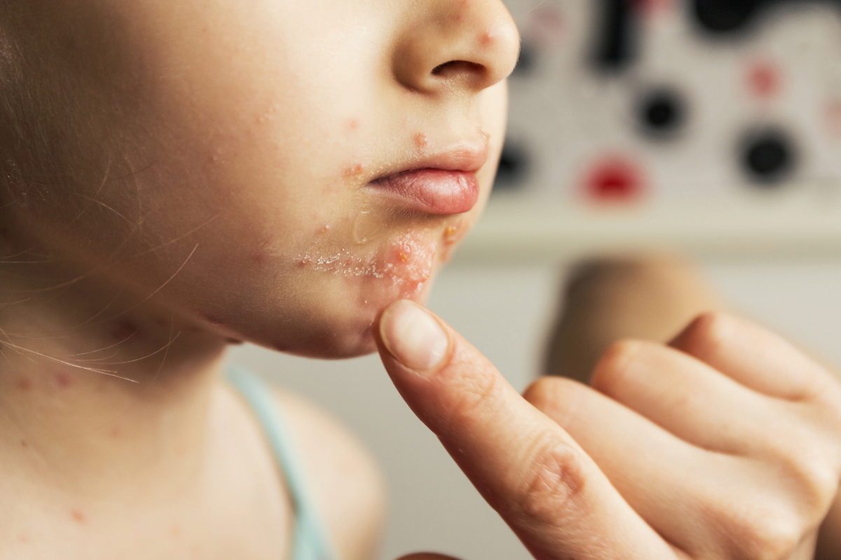
  • Dyshidrotic Eczema and Autoimmune Disease: Symptoms and Care

    Dyshidrotic Eczema and Autoimmune Disease: Symptoms and Care

    Dyshidrotic eczema and autoimmune disease are related through immune activity, but dyshidrotic eczema is not usually classified as a classic autoimmune disease. It is an inflammatory skin condition that causes deep, itchy blisters on the hands or feet. The distinction matters because treatment often focuses on calming inflammation, repairing the skin barrier, and finding triggers rather than treating a systemic autoimmune illness.

    Some people also live with allergies, atopic dermatitis, psoriasis, lupus, or other immune-related conditions. That overlap can make flares feel harder to interpret. This practical overview explains what the blisters mean, when immune-system questions deserve follow-up, and how to discuss diagnosis and care with a clinician.

    Key Takeaways

    • Not usually autoimmune: Dyshidrotic eczema is generally considered inflammatory, not a classic autoimmune disease.
    • Blisters have patterns: Deep, itchy vesicles often affect palms, fingers, soles, and toes.
    • Triggers are personal: Sweat, stress, metals, irritants, allergies, and wet work can contribute.
    • Diagnosis is clinical: Exams, fungal testing, patch testing, or biopsy may help rule out lookalikes.
    • Care is layered: Barrier repair, trigger control, topical medicines, phototherapy, and systemic options may be considered.

    Is Dyshidrotic Eczema Autoimmune?

    Dyshidrotic eczema is best understood as an immune-influenced inflammatory skin disorder. In autoimmune disease, the immune system targets the body’s own tissues in a more specific way. Dyshidrotic eczema, also called pompholyx eczema, appears to involve immune overactivity, barrier disruption, and trigger sensitivity, but it is not usually grouped with conditions like lupus or rheumatoid arthritis.

    That said, the immune system still plays a real role. During a flare, inflammatory signals can make the skin swell, itch, and form tiny fluid-filled blisters. People with allergic tendencies, chronic hand eczema, or other inflammatory skin conditions may be more prone to repeated flares.

    Why it matters: Knowing the difference helps you ask better questions and avoid assuming every flare means systemic autoimmune disease.

    If you want a broader framework for immune conditions, Autoimmune Diseases explains general terminology and patterns. For skin-specific comparisons, the Dermatology category can help you browse related education.

    What the Blisters Look and Feel Like

    Dyshidrotic eczema symptoms usually appear as small, deep-seated blisters on the palms, sides of the fingers, soles, or toes. These blisters, also called vesicles, may feel intensely itchy before they become visible. Some people describe a burning or prickling sensation under the skin.

    Flares often come in waves. Blisters can cluster, merge, dry out, peel, and leave cracks behind. When fissures form, daily tasks such as typing, cooking, washing dishes, walking, or wearing shoes may become painful. Between flares, the skin may remain dry, thickened, scaly, or sensitive to soaps and friction.

    Dyshidrotic eczema hands involvement can look like fingertip peeling, split cuticles, or blisters along the sides of the fingers. Dyshidrotic eczema feet symptoms may affect the arches, sides of the feet, or toes. Foot flares can be mistaken for athlete’s foot, especially when scaling or itching is prominent.

    Pictures can support pattern recognition, but they cannot confirm the diagnosis. Several palm-and-sole rashes can look similar. If pustules, thick plaques, or persistent scaling dominate, Pustulosis vs Dyshidrotic Eczema outlines important differences to discuss with a clinician.

    Why Flares Happen Suddenly

    A sudden dyshidrotic eczema flare up often reflects several factors stacking together. A person may have a fragile skin barrier, then encounter sweat, friction, metal exposure, frequent washing, or emotional stress. The immune system may then react strongly, producing itching and blisters.

    Common dyshidrotic eczema triggers include nickel, cobalt, fragrances, detergents, hand sanitizers, rubber additives, and harsh cleansers. Wet work is another major issue. This includes repeated washing, cleaning, food preparation, healthcare work, hairdressing, and wearing occlusive gloves for long periods. Sweat trapped inside gloves or shoes can worsen irritation.

    Stress does not mean a flare is “all in your head.” Stress hormones can influence inflammation, itch perception, sweating, and sleep. If stress appears to line up with outbreaks, Stress and Dyshidrotic Eczema offers a deeper look at that connection.

    Some people ask about the “rule of 3” for eczema. In everyday skin care, it is often used as a simple habit reminder rather than a formal medical rule: moisturize often, avoid known triggers, and treat flares early according to your care plan. Your clinician may use different wording, but the idea is consistent: protect the barrier before the flare becomes severe.

    Immune Links, Lupus Questions, and Comorbid Conditions

    Dyshidrotic eczema and autoimmune disease can overlap in a person’s health history without one proving the other. For example, someone may have dyshidrotic eczema and also have lupus, thyroid disease, or another immune-mediated condition. That does not mean the blisters are caused by the autoimmune diagnosis in every case.

    Lupus deserves special care because it can cause several skin findings, including photosensitive rashes and lesions that may need different evaluation. Dyshidrotic eczema lupus concerns are worth raising if you also have joint swelling, mouth ulcers, unusual sun-sensitive rashes, fevers, chest pain, or unexplained fatigue. These symptoms do not confirm lupus, but they can justify a broader medical review.

    Allergic contact dermatitis is another common overlap. It happens when the immune system reacts to a substance touching the skin. Nickel, cobalt, fragrance, preservatives, and rubber chemicals can all be relevant. Patch testing can help identify delayed contact allergies that keep hand or foot eczema active.

    Atopic dermatitis, psoriasis, and palmoplantar pustulosis may also enter the discussion. Some treatments and prevention steps overlap, but diagnosis matters. A treatment plan for fungal infection or psoriasis may differ from one for pompholyx eczema, so persistent or unusual symptoms should not be self-labeled for long.

    Diagnosis: What Clinicians Usually Check

    Dyshidrotic eczema diagnosis usually starts with an exam and symptom history. A clinician looks at where the blisters appear, how deep they are, whether the skin peels or cracks, and whether both sides of the body are involved. Timing also matters. Repeated flares after sweat, wet work, metal exposure, or stress can provide clues.

    A potassium hydroxide test, often called a KOH test, may be used when fungal infection is possible. This is especially relevant for foot symptoms. Patch testing may be suggested when contact allergy is suspected or flares keep returning despite basic skin care. Rarely, a skin biopsy may help rule out other inflammatory or blistering disorders.

    Prepare for your appointment by noting patterns. Track when flares start, where they appear, what products touched the skin, and whether new gloves, shoes, hobbies, or workplace exposures changed recently. Photos from early blister stages can help, because lesions may look different by the appointment date.

    Quick tip: Bring ingredient lists from soaps, creams, gloves, and workplace products when contact allergy is possible.

    Treatment Options and Home Care Steps

    Dyshidrotic eczema treatment aims to reduce inflammation, repair the barrier, manage itch, and prevent infection. The right plan depends on severity, location, infection risk, pregnancy status, other conditions, and past response to treatments. A clinician can help match treatment intensity to the flare.

    Barrier repair and prevention

    Daily barrier care is the foundation. Fragrance-free moisturizers, petrolatum-based ointments, and ceramide-containing creams can reduce dryness and cracking. Apply moisturizer after washing and before activities that expose your skin to friction. Cotton gloves at night may help keep ointment in place, but they should not trap moisture during active sweating.

    For wet work, try to reduce repeated soap exposure when possible. Use lukewarm water rather than hot water. Choose mild cleansers, rinse well, and moisturize quickly. If gloves are needed, cotton liners under nitrile gloves may reduce sweat contact. Change damp liners instead of wearing them for long stretches.

    Prescription and clinic-based options

    During flares, clinicians may recommend topical corticosteroids for short periods, especially on thick palm or sole skin. Potency and duration matter because overuse can increase side effects. If a potent steroid is prescribed, Clobetasol Side Effects explains monitoring points to discuss.

    Non-steroidal topical immunomodulators may be considered in some eczema care plans. Product pages such as Protopic Ointment and Tacrolimus HGC can provide medication-specific context, but treatment choices should come from your clinician. For more severe inflammatory skin disease, systemic or biologic therapies may be reviewed. Dupixent Explained offers general background on one biologic option used in certain eczema-related care contexts.

    Other options may include phototherapy, wet dressings, antihistamines for sleep-disrupting itch, infection treatment when needed, or systemic anti-inflammatory medicines for severe disease. Do not start, stop, or change prescription treatments without medical guidance.

    When Dyshidrotic Eczema Can Become Risky

    Dyshidrotic eczema is not contagious, but cracked or open skin can become infected. You cannot spread dyshidrotic eczema to another person through touch, towels, or shared surfaces. Infection risk comes from damaged skin, not from the eczema itself being a germ.

    Seek medical care promptly if you notice pus, honey-colored crusting, spreading redness, warmth, fever, red streaks, or rapidly worsening pain. People with diabetes, circulation problems, immune suppression, or frequent foot wounds should be especially cautious. Foot fissures can affect walking and may need earlier evaluation.

    Function is also a safety issue. If hand pain prevents work, sleep, hygiene, or basic self-care, it is reasonable to ask whether your current plan is strong enough. Occupational exposures may require practical adjustments, protective equipment, or a clinician note when appropriate.

    Everyday Prevention That Respects Real Life

    Dyshidrotic eczema prevention works best when it is realistic. You do not need a perfect routine. You need a repeatable one that lowers irritation while supporting work, caregiving, hobbies, and sleep.

    • Track flare timing: Note sweat, stress, products, foods, work tasks, and weather shifts.
    • Simplify products: Choose fragrance-free cleansers, moisturizers, and laundry products when possible.
    • Protect during wet work: Use gloves thoughtfully, and replace damp liners quickly.
    • Reduce friction: Rotate shoes, wear breathable socks, and avoid tight footwear during foot flares.
    • Ask about patch testing: Consider this when flares keep returning or exposures are unclear.
    • Follow up early: Escalate care when blisters crack, spread, or disrupt daily function.

    Some people also review diet when metal allergy is confirmed, especially nickel sensitivity. Dietary changes should be discussed with a clinician or dietitian rather than started broadly, because unnecessary restriction can make eating harder without clear benefit.

    For readers comparing products or prescription categories, the Dermatology Products category may help with general navigation. Keep product research separate from diagnosis, since a similar-looking rash may need different treatment.

    Authoritative Sources

    For patient-focused self-care steps and treatment context, review the American Academy of Dermatology dyshidrotic eczema guidance. It covers gentle skin care, flare management, and when dermatology input may help.

    For a patient-education overview of triggers, symptoms, and treatment categories, see the National Eczema Association dyshidrotic eczema resource. It is useful for understanding common patterns and care conversations.

    For a research-focused discussion of immune mechanisms, the immune response in pompholyx review summarizes how local immune findings have been studied. Research language can be technical, so use it as background rather than personal diagnosis.

    Putting the Pieces Together

    Dyshidrotic eczema and autoimmune disease can feel connected because both involve the immune system. The more accurate view is that dyshidrotic eczema is usually an inflammatory, trigger-sensitive skin condition rather than a classic autoimmune disease. Still, immune history matters when symptoms are severe, unusual, widespread, or paired with systemic signs.

    What to do next depends on your pattern. Track triggers, protect the skin barrier, ask about contact allergy, and seek evaluation when symptoms do not fit the usual course. If you have joint swelling, photosensitive rashes, fevers, or other whole-body symptoms, raise those concerns directly with your healthcare professional.

    This content is for informational purposes only and is not a substitute for professional medical advice.

  • Stress Dyshidrotic Eczema: Role, Triggers, and Contagious Risk

    Stress Dyshidrotic Eczema: Role, Triggers, and Contagious Risk

    If tiny, itchy hand or foot blisters keep returning, stress may be part of the picture. Many people call this pompholyx or vesicular hand eczema; clinicians may say dyshidrotic eczema. We review stress biology, everyday triggers, and safe care options. You’ll also learn whether stress dyshidrotic eczema spreads and what to do next.

    Key Takeaways

    • Stress link: Nervous system changes can intensify itch and blisters.
    • Not infectious: Fluid-filled blisters do not spread to others.
    • Trigger stacking: Metals, sweat, heat, and friction often combine.
    • Gentle care: Emollients, cool soaks, and trigger control help.
    • Medical support: Topicals and light therapy may calm stubborn flares.

    Stress Dyshidrotic Eczema

    Stress activates the brain–skin axis, which can intensify itch, sweat, and inflammation. When stress hormones rise, skin barrier function may weaken, and nerve-driven itch can spiral. Scratching then releases more inflammatory signals, creating a flare loop. In dyshidrotic eczema (also called pompholyx), this loop shows up as clusters of deep, tapioca-like blisters on palms, fingers, or soles.

    Clinicians describe this pattern as a chronic, relapsing form of vesicular hand eczema. People often report flares during exams, shift changes, or family stress. Heat, occlusive gloves, or sudden sweating can amplify symptoms. Addressing stress alongside skin care breaks the cycle more effectively than either strategy alone.

    What It Is and What It Isn’t

    This condition features intensely itchy, fluid-filled vesicles on hands and feet that may coalesce into plaques. When the blisters dry, the skin can crack and peel, causing pain and reduced grip. Typical Eczema patterns help clinicians recognize the condition. For a broader baseline definition, see Eczema as a starting point.

    Common dyshidrotic eczema symptoms include tingling, burning, and visible vesicles on the sides of fingers, palms, and soles. Unlike infections, there is no contagious organism. Unlike psoriasis, there are no thick silvery scales. And unlike scabies, burrows are absent. Confusion with allergic contact dermatitis is possible, so history and exposure review matter.

    Triggers and Risk Factors

    Triggers are highly individual, yet some patterns repeat. Nickel exposure from jewelry, keys, or coins may provoke flares, as can cobalt, fragrance, and harsh detergents. Heat and occlusion trap moisture and irritants against the skin. Many people note seasonal allergies or atopic tendencies in their history. The cumulative burden of small triggers often exceeds any single cause.

    Because trigger stacking is common, tracking patterns guides prevention. People often report dyshidrotic eczema triggers like sweaty palms during deadlines, vigorous workouts, or prolonged glove use. For background on stress physiology and its body-wide effects, see Science of Stress, which explains why stress can magnify itch and inflammation. Those with an atopic history may also benefit from Atopic Dermatitis Eczema for context on allergic tendencies.

    Diagnosis and When to Seek Care

    Diagnosis is usually clinical, based on the blisters’ depth, distribution, and a relapsing course. Your clinician may ask about metal exposure, occupational irritants, and family history of atopy. If fungal infection is suspected, they may perform a skin scraping to rule out tinea. Select patch testing can identify metal or fragrance allergy when contact reactions are suspected.

    If blisters are severe, painful, or disrupt work and sleep, seek evaluation. Discuss recent exposures, new skincare products, and glove or gym routines. When patterns are unclear, asking about dyshidrotic eczema diagnosis steps—like patch testing for nickel or cobalt—can help. For a side-by-side view of overlapping rashes, see Palmoplantar Pustulosis vs. Dyshidrotic Eczema to understand key differences and avoid confusion.

    Contagious or Not? Understanding Spread

    These blisters are not contagious. The clear fluid is sterile, not infectious pus. You cannot give it to someone else through touch, towels, or shared surfaces. This is a noninfectious inflammatory process tied to skin barrier function, nerves, sweat, and immune responses. The term dyshidrosis is sometimes used interchangeably, reflecting the characteristic vesicles on hands and feet.

    Authoritative dermatology sources consistently note the noninfectious nature of this rash. According to the American Academy of Dermatology, dyshidrotic eczema is a type of eczema and does not spread from person to person. For concise clinical descriptions of presentation and triggers, see DermNet, which provides accessible summaries for patients and clinicians.

    Managing Flares and Daily Care

    Calming the skin and protecting the barrier are daily priorities. Use a thick, fragrance-free emollient several times a day. Ointments containing petrolatum can reduce transepidermal water loss. During itchy episodes, cool compresses or diluted astringent soaks (such as aluminum acetate) may provide short-term relief. Patch test new products before widespread use.

    Simple routines often help more than complex ones. Wear breathable cotton liners under work gloves, then moisturize after removal. Keep nails short to reduce scratch injury. Gentle handwashing with lukewarm water, followed by immediate emollient application, supports barrier healing. People often search for dyshidrotic eczema home remedies like diluted vinegar soaks; discuss any regimen with a clinician if your skin is cracked, infected, or very painful. For background terminology and subtypes, see Eczema Dermatitis, which outlines broader dermatitis categories.

    Medical Treatments and Evidence

    Topical anti-inflammatory agents are the mainstay for localized flares. Potent topical corticosteroids may be used short term on palms and soles, with careful monitoring for side effects. Nonsteroidal options like calcineurin inhibitors (tacrolimus, pimecrolimus) can help maintenance on thin skin areas or when steroid-sparing is preferred. Phototherapy may be considered for frequent relapses or widespread involvement.

    Ask your clinician about dyshidrotic eczema treatment choices that fit your pattern and medical history. For an overview of a nonsteroidal option, see Protopic Ointment, referenced here because tacrolimus is sometimes used in sensitive areas. For high-potency steroid discussions, Clobetasol is linked as an example of a class option; potency and duration should always be clinician-directed. For safety considerations and adverse effects, consult Clobetasol Propionate Side Effects to understand risk discussions before use.

    Stress Skills and Relapse Prevention

    Addressing stress biology is not optional; it is part of care. Brief, regular practices can reduce itch-scratch cycles and improve sleep. Evidence-informed approaches include paced breathing, muscle relaxation, guided imagery, and mindfulness. Short walk breaks, consistent bedtimes, and phone-free wind-down routines can lower physiologic arousal. Consider scheduling micro-breaks before high-demand tasks to prevent over-activation.

    Personalize your plan and keep it simple. Some people find cognitive behavioral strategies helpful for cue-based scratching. Others prefer yoga, tai chi, or quiet reading. Building a small daily habit is better than occasional intensity. Integrating stress management for eczema with barrier care reduces flare severity for many. If flares persist despite strong self-care, ask your clinician about allergy testing or referral.

    When Nickel and Sweat Intersect

    Metal allergy and perspiration often interact. Sweat can mobilize nickel ions from coins, keys, or accessories, increasing skin exposure. Moist hands under gloves or during workouts can magnify contact effects. Practical steps include swapping to low-nickel gear and drying hands thoroughly before glove use. Rotating tasks to limit prolonged occlusion also helps.

    If you suspect nickel sensitivity, consider discussing patch testing and exposure reduction with your clinician. Switching to nonmetal accessories, key covers, and compatible watchbands can make a difference. Keep in mind that dietary nickel is a separate topic; dietary changes should be guided by a clinician or dietitian. People with persistent blisters and suspected metal allergy sometimes ask about “best creams”; focus first on trigger control, then individualized medications.

    Safety, Complications, and Infections

    Although extremely itchy and disruptive, this rash is typically not dangerous. The main risks involve skin cracks, sleep loss, and secondary infection. Seek care if you notice spreading redness, pus, fevers, or severe pain. Clinicians may culture suspicious lesions or prescribe antibiotics if infection is confirmed. Hand function at work can also suffer, so documentation may help workplace accommodations.

    Protecting cracked skin reduces complications. Cover splits with occlusive dressings after emollients. Consider cotton glove liners for chores and replace harsh cleansers with gentle options. If over-the-counter products sting or worsen the rash, stop and ask for guidance. People often try remedies they read online; it’s wise to discuss any new regimen with a clinician who knows your skin history.

    Prevention in Daily Life

    Consistency wins. Moisturize after every handwash and before bed. Keep a small tube of ointment in your bag, car, or desk. Swap fragranced soaps for gentle, fragrance-free cleansers. Choose breathable gloves for tasks, then moisturize immediately afterward. Plan ahead for heat and sweat by carrying a clean towel and scheduling brief cool-downs.

    Review exposures weekly and adjust one variable at a time. Replace metal-touch habits when possible, and track flare timing in a simple note app. If you see patterns tied to work, discuss protective options with your supervisor. Small environmental changes often add up to fewer flares over time. When uncertainty remains, re-evaluate triggers and ask about patch testing to clarify hidden culprits.

    Note: If symptoms change suddenly or involve severe pain, swelling, or signs of infection, seek prompt evaluation.

    This content is for informational purposes only and is not a substitute for professional medical advice.

  • Chickenpox vs Shingles: Symptoms, Rash, and Contagion

    Chickenpox vs Shingles: Symptoms, Rash, and Contagion

    Chickenpox vs shingles comes down to timing, rash pattern, and how the virus spreads. Chickenpox is usually the first infection with varicella-zoster virus. Shingles, also called herpes zoster, happens when that same virus reactivates later. Chickenpox often causes an itchy, widespread rash. Shingles more often causes pain, burning, or tingling followed by a one-sided band or cluster of blisters. That difference matters because exposure risk, isolation needs, vaccine history, and urgency of care can vary.

    Key Takeaways

    • Same virus: Both conditions involve varicella-zoster virus.
    • Different timing: Chickenpox is usually first infection; shingles is reactivation.
    • Rash clues: Chickenpox spreads widely; shingles often stays one-sided.
    • Contagion differs: Shingles can expose non-immune people to chickenpox.
    • Seek care quickly: Eye symptoms, pregnancy, newborn exposure, and weak immunity matter.

    Chickenpox vs Shingles: The Core Difference

    The key difference is that chickenpox usually starts the infection, while shingles is the virus waking up later. After chickenpox, varicella-zoster virus can stay inactive in nerve tissue for years. It may reactivate when immune defenses change with age, illness, stress, or immune-suppressing treatment.

    This explains a common point of confusion. A person does not usually “catch shingles” from someone else in the same way they catch a cold. Shingles is usually caused by a virus already inside the body. However, fluid from shingles blisters can spread varicella-zoster virus to someone without immunity. That exposed person may develop chickenpox, not shingles, because it would be their first infection.

    The medical names can also confuse people. Varicella usually refers to chickenpox. Herpes zoster refers to shingles. The word “herpes” here does not mean herpes simplex virus, which causes cold sores and genital herpes. If you want broader context on herpes simplex symptoms, see Herpes Symptoms.

    Comparison point Chickenpox Shingles
    Usual cause First varicella-zoster virus infection Reactivation of varicella-zoster virus
    Typical rash Widespread itchy spots and blisters Painful cluster or band, often one-sided
    Early symptoms Fever, tiredness, appetite loss, then rash Burning, tingling, pain, then rash
    Contagion Spreads easily to susceptible people Spreads mainly through blister fluid contact
    Prevention focus Varicella vaccination Shingles vaccination for eligible adults

    Why it matters: Similar blisters can mean different risks for nearby people.

    How the Rash and Early Symptoms Usually Differ

    Symptoms often provide the first clue before the rash fully appears. Chickenpox tends to act like a whole-body viral illness. Shingles often starts with nerve-type symptoms in one limited area.

    Early chickenpox symptoms

    Chickenpox may begin with fever, tiredness, headache, appetite loss, or a general unwell feeling. The rash often starts as red spots that become fluid-filled blisters. New spots may appear in waves, so some lesions can be new while others are crusting.

    The itch can be intense. Scratching may break the skin and raise the chance of bacterial infection. Children often have milder illness than adults, but severity can vary. Adults, newborns, pregnant people, and people with weakened immune systems may face higher risk from chickenpox.

    First signs of shingles

    Shingles may start with pain, burning, tingling, itching, or unusual skin sensitivity. This early warning phase is called a prodrome, which means symptoms before the main illness becomes obvious. The discomfort may feel sharp, electric, deep, or tender to touch.

    The rash usually appears in a localized strip, patch, or cluster. It often stays on one side of the body and follows a nerve pathway. A classic example is a band of blisters wrapping around one side of the torso. Shingles can also affect the face, scalp, mouth, or eye area.

    A rash near the eye, ear, or face deserves prompt medical attention. Shingles in these areas can involve sensitive structures, and early evaluation may help reduce complications.

    Can Shingles Cause Chickenpox or Spread to Others?

    Yes, shingles can expose a susceptible person to varicella-zoster virus. If that person never had chickenpox, never received varicella vaccination, and has no immunity, they may develop chickenpox after exposure. They would not simply develop shingles from casual contact.

    Chickenpox is usually more contagious than localized shingles. It can spread through respiratory droplets and direct contact with lesions, especially before every spot has crusted. Shingles spreads mainly through direct contact with fluid from open blisters. Covered, crusted lesions carry less practical contact risk, though individual circumstances still matter.

    Questions about kissing, hugging, or sharing a bed depend on rash location and immunity status. If shingles blisters are on the face or near the mouth, kissing may create direct contact with blister fluid. If the rash is covered and away from contact areas, risk may be lower, but vulnerable contacts still need caution.

    Avoid touching the rash. Keep it covered when possible. Do not share towels, bedding, or clothing that may contact blister fluid. People with active shingles should be especially cautious around newborns, pregnant people without known immunity, and people who are immunocompromised, meaning their immune system is weakened.

    Quick tip: If exposure is possible, write down the rash date and contact date.

    Can You Get Shingles If You Never Had Chickenpox?

    Shingles usually requires prior varicella-zoster infection or prior exposure through vaccination. If someone truly never had chickenpox, never had varicella vaccination, and never had an unnoticed infection, they would not be expected to develop shingles from reactivation. In real life, many people do not remember childhood infections accurately.

    A clinician may review vaccine records, exposure history, or blood testing when immunity is unclear. This can matter for pregnancy, healthcare work, immune-suppressing treatment, or close contact with high-risk people. It also helps explain why “I never had chickenpox” does not always settle the question.

    Chickenpox vs shingles can be especially confusing in adults. Adults may have more severe chickenpox than children. Shingles becomes more common with age, but it can occur in younger adults too. A painful one-sided rash should not be dismissed just because someone is not elderly.

    Treatment, Comfort Care, and Vaccine Context

    Treatment depends on the person, timing, symptoms, and complication risk. Chickenpox care is often supportive. That may include fluids, rest, fever comfort measures, and itch control. Clinicians generally caution against aspirin in children with viral illnesses because of Reye’s syndrome risk.

    Shingles care may include prescription antiviral medicine for some people, along with pain control and skin care. Early assessment matters because antiviral treatment can be time-sensitive in higher-risk cases. Do not start, stop, or change prescription treatment without professional guidance.

    Practical comfort measures may include:

    • Skin protection: Keep fingernails short.
    • Rash hygiene: Keep lesions clean and dry.
    • Contact control: Cover shingles blisters when possible.
    • Symptom notes: Track fever, pain, and rash spread.
    • Medication review: Ask what is safe for your age and history.

    Antibiotics do not treat chickenpox or shingles because both involve a virus. A clinician may consider antibiotics only if a bacterial skin infection develops. That decision requires an exam and clinical judgment.

    Vaccination is another major difference. Varicella vaccination is used to help prevent chickenpox. Shingles vaccination is aimed at reducing the risk of shingles and related complications in eligible adults. Recommendations can depend on age, immune status, prior infection, and local public health guidance.

    For readers comparing skin infections, the Dermatology collection and Infectious Disease collection can support broader reading. These pages are for navigation and education, not self-diagnosis.

    When a prescription is required, licensed pharmacy partners may verify prescription details with the prescriber before dispensing. Some eligible patients also explore cash-pay cross-border prescription options without insurance, subject to local rules and clinical requirements.

    When Chickenpox or Shingles Is More Concerning

    Neither condition should be treated as “just a rash” in higher-risk situations. Most healthy children recover from chickenpox without major problems, but complications can occur. Adults with chickenpox may have more severe illness than children. Infants, pregnant people, and people with weakened immune systems need careful assessment.

    Shingles can also become complicated. Some people develop postherpetic neuralgia, which is nerve pain that persists after the rash heals. Shingles near the eye can threaten vision. Widespread shingles, severe pain, fever, confusion, or a rash in someone with a weakened immune system may need urgent evaluation.

    So, which is worse: chickenpox or shingles? The safer answer is that it depends. Chickenpox can be more concerning for adults, pregnancy, newborn exposure, and people without immunity. Shingles can be especially concerning when pain is severe, the face or eye is involved, or immune defenses are reduced.

    Vitamin deficiency is sometimes discussed online in relation to shingles. Low nutrition status can affect general immune health, but shingles is not diagnosed as a simple vitamin-lack problem. If fatigue, poor intake, or repeated infections are concerns, a clinician can assess the broader picture.

    Look-Alike Rashes: Herpes, Measles, and Other Causes

    Several infections and skin conditions can mimic parts of chickenpox or shingles. Herpes simplex can cause painful blisters, often around the mouth or genitals, but it is caused by a different virus. A localized painful blistering rash may need testing or clinical review when the cause is unclear.

    Measles can cause fever and a widespread rash, yet it usually has additional respiratory symptoms and a different illness pattern. Allergic reactions, insect bites, contact dermatitis, impetigo, and drug reactions can also create confusing rashes. Appearance alone is not always enough.

    Recurrent chickenpox is uncommon. When someone thinks chickenpox has returned, shingles, herpes simplex, allergic reactions, or other rashes may be part of the differential diagnosis. For related viral-rash reading, Herpes Treatment discusses herpes simplex management concepts separately from varicella-zoster illness.

    Do not rely on photos alone if the rash is painful, near the eye, spreading quickly, or affecting someone at higher risk. Photos can document changes, but they cannot replace a medical exam when warning signs are present.

    A Practical Checklist Before You Call a Clinician

    A clear history can make the visit more useful. Before calling or attending an appointment, gather details that help separate first infection, reactivation, and look-alike rashes.

    • Rash timing: Note when pain, itch, or spots began.
    • Rash location: Record whether it is one-sided or widespread.
    • Exposure history: List known chickenpox or shingles contact.
    • Vaccine record: Check varicella and shingles vaccine history.
    • Risk factors: Mention pregnancy or immune conditions.
    • Eye symptoms: Report redness, pain, or vision changes.
    • Medication list: Include prescriptions, supplements, and allergies.

    This information does not diagnose the rash by itself. It helps your healthcare professional decide what questions, tests, or precautions may be appropriate.

    Authoritative Sources

    Putting the Difference Into Practice

    Chickenpox vs shingles is easiest to understand by asking four questions. Is this the first infection or a reactivation? Is the rash widespread or one-sided? Is itching or pain the main symptom? Could someone nearby be vulnerable because of pregnancy, newborn age, or weakened immunity?

    A widespread itchy rash points more toward chickenpox. A painful one-sided cluster points more toward shingles. If symptoms involve the eye, pregnancy, a newborn, severe pain, fever, or a weakened immune system, seek medical guidance promptly.

    This content is for informational purposes only and is not a substitute for professional medical advice.